Trichoscopy of inflammatory conditions — extra information
Extra information
Synonyms:
Dermoscopy of inflammatory conditions, Trichoscopy of psoriasis, Trichoscopy of dermatomyositis, Trichoscopy of seborrhoeic dermatitis, Trichoscopy of scalp psoriasis, Trichoscopy of systemic sclerosis
Author: Dr Ahmed Sadek, Cairo Hospital for Dermatology & Venereology (Al-Haud Al-Marsoud), Egypt. June 2022.
Contributors: Dr Dalia Hossam, Dr Radwa Magdy, Dr Nehal Saied, Dr Noha Hashem, Dr Safaa Yehia Negm, Dr Moshera Saied El Bahrawy, Dr Amira Ragab, Dr Amal Wagih, Dr Haidy El-Hussieny, Dr Mona Ragib, Dr Hala Amer. Copy edited by Gus Mitchell. June 2022
The following page covers the features of inflammatory skin disease using trichoscopy.
Seborrheic dermatitis
Seborrheic dermatitis is a common chronic relapsing inflammatory skin condition with a predilection for areas rich in sebaceousglands (i.e. the scalp, central face, and anterior chest are most affected). The disorder is characterised by scaling with poorly defined erythematous patches and plaques. Young adults are more frequently affected, and men are affected more than women.
Multiple, thin, arborizing vessels are observed; recently, comma vessels have also been reported.
Featureless areas with no particular vascular pattern may be found
Adherent yellow scales and interfollicular white scales
Interfollicular oily material
Interfollicular pustules
Honeycomb pigment pattern
Concretions
Yellow dots
“Dandelion sign” described as yellow dot surrounded by glomerular and comma vessels
“Cherry blossom sign” described as arborizing vessels with glomerular and comma vessels surrounding them.
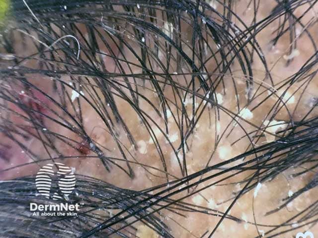
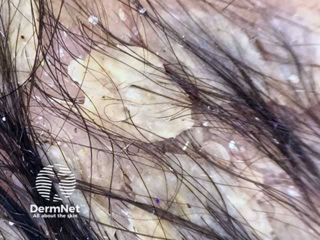
Dermoscopic image of diffuse yellowish greasy scales in a male patient with seborrhoeicdermatitis
Dermoscopic image of seborrhoeic dermatitis showing adherent yellowish scales and interfollicular oily material on the scalp of a male infant (SD-patient1)
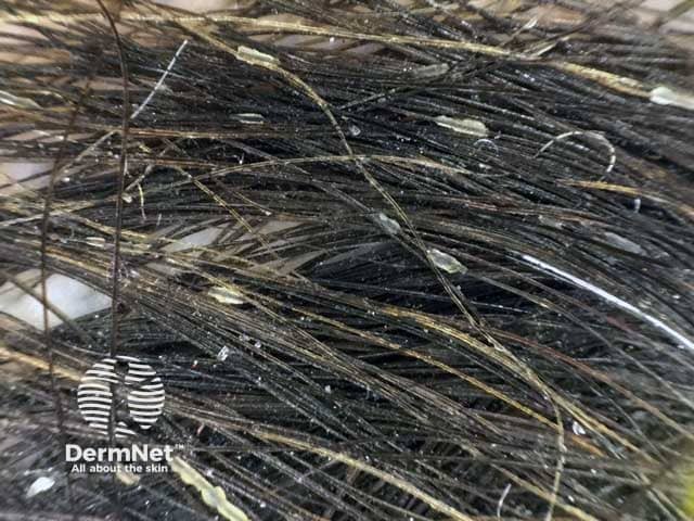
Dermoscopic image of hair casts represented by detached hyperkeratotic and parakeratotic cells of the inner root sheath which surrounds the hair shaft
Psoriasis
Psoriasis is a chronic inflammatory cutaneous disorder with multisystem involvement. The scalp is the most commonly affected site (50–80% of psoriasis patients) and may be the first or only disease manifestation.
Scalp psoriasis is characterised by sharply bordered, erythematous lesions covered by silver or white scales. The lesions often spread over the anterior hairline into the forehead and may also affect the retro-auricular region.
Trichoscopic evaluation of scalp psoriasis
Evaluation of scalp psoriasis is based on the evaluation of vascular alterations.
At low magnifications:
Red dots and globules with regular distribution are observed
White/white-silver dry scales is a very frequent finding
Hidden hair and signet ring vessels are recently reported signs supporting the diagnosis of psoriasis.
At high magnifications:
Glomerular vessels arranged into rings or lines
Other types of capillaries (linear looped, lace-like, comma vessels) may be present.
Systemic sclerosis (SSc)
Systemic sclerosis is an autoimmuneconnective tissue disease. Different factors are incorporated in the pathogenesis of the disease including; small vessel vasculopathy, production of autoantibodies, and fibroblastdysfunction.
Clinical manifestations and prognosis are variable according to the degree of cutaneous and systemic organs involvement. Clinical subtypes include limited cutaneous SSc, diffuse cutaneous SSc, and SSc without skin involvement.
Polymorphous pattern of blood vessels, including arborizing vessels similar to the pattern observed in DLE, spider vessels, and capillary loops
Telangiectasia detected in the scalp of systemic sclerosis patients was also observed in other sites of the body
‘Salt and pepper’ areas are a recently reported sign
Loss of follicular openings
Pili torti
Black dots
Broken hairs.
Dermatomyositis
Dermatomyositis is a chronic inflammatory disease characterised by proximal myopathy, and cutaneous, cardiac, pulmonary, and gastrointestinal affection. Common cutaneous signs include Gottron's papules, heliotrope rash, mechanic's hand, and poikiloderma.
Trichoscopic findings
Erythematous violaceous areas.
Enlarged tortuous and bushy capillaries.
Interfollicular scales.
Interfollicular and perifollicularpigmentation.
Scalp atrophy.
Tufting with three or more hair shafts emerging together surrounded by peripilar cast.
Bibliography
Golińska J, Sar-Pomian M, Rudnicka L. Dermoscopic features of psoriasis of the skin, scalp and nails - a systematic review. J Eur Acad Dermatol Venereol. 2019;33(4):648–60. doi:10.1111/jdv.15344. Journal
Jasso-Olivares JC, Tosti A, Miteva M, Domínguez-Cherit J, Díaz-González JM. Clinical and Dermoscopic Features of the Scalp in 31 Patients with Dermatomyositis. Skin Appendage Disord. 2017;3(3):119–24. doi:10.1159/000464469. Journal
Kibar M, Aktan Ş, Bilgin M. Dermoscopic findings in scalp psoriasis and seborrheic dermatitis; two new signs; signet ring vessel and hidden hair. Indian J Dermatol. 2015;60(1):41–5. doi:10.4103/0019-5154.147786 Journal
Kim GW, Jung HJ, Ko HC, et al. Dermoscopy can be useful in differentiating scalp psoriasis from seborrhoeic dermatitis. Br J Dermatol. 2011;164(3):652–6. doi:10.1111/j.1365-2133.2010.10180.x. Journal
Kłosowicz A, Alsalhi W, Tosti A. How to Optimize Trichoscopy for Evaluation of Scalp Vessels. Skin Appendage Disord. 2020;6(4):216–9. doi:10.1159/000508166. Journal
Kwiatkowska M, Rakowska A, Walecka I, Rudnicka L. The diagnostic value of trichoscopy in systemic sclerosis. J Dermatol Case Rep. 2016;10(2):21–5. Published 2016 Nov 13. doi:10.3315/jdcr.2016.1225. PubMed Central
Ruiz-Arriaga LF, Arenas R, Vega-Sánchez DC, Asz-Sigall D, Martínez-Velazco MA. Seborrheic Dermatitis: Three Novel Trichoscopic Signs and Its Correlation to Malassezia sp. Colonization. Skin Appendage Disord. 2019;5(5):288–92. doi:10.1159/000497782. Journal
Widaty S, Pusponegoro EH, Rahmayunita G, et al. Applicability of Trichoscopy in Scalp Seborrheic Dermatitis. Int J Trichology. 2019;11(2):43–8. doi:10.4103/ijt.ijt_86_18. Journal