Authors: Vanessa Ngan, Staff Writer, 2003. Dr Sabrina Sapsford, Clinical Medical Education Fellow, University of Auckland, Auckland, New Zealand. Copy edited by Gus Mitchell. October 2020.
Dermatomyositis is an idiopathicinflammatorymyopathy characterised by skeletal muscle weakness and skin changes.
Who gets dermatomyositis?
Dermatomyositis is uncommon, with an annual incidence of 0.1-6 per population of 100,000. It can affect people of any race, age, and sex; however, women are affected twice as often as men, and Black Americans are more commonly affected than white Americans. It can present in children (see Juvenile dermatomyositis). The peak age group affected in adults is those aged 50–60 years.
Adult-onset dermatomyositis is strongly associated with malignancy; up to 25% of affected adults have an unknown underlying malignancy on diagnosis. The majority are adenocarcinomas.
What causes dermatomyositis in adults?
Dermatomyositis is thought to be caused by a microangiopathy affecting skin and muscle. There is a geneticpredisposition to the development of dermatomyositis such as the PTPN22 gene and HLA associations identified include:
Caucasian populations: HLA DRB1*0301 and DQA1*0501
Most patients have disease-associated autoantibodies, suggesting dermatomyositis is an autoimmune condition.
What are the clinical features of adult-onset dermatomyositis?
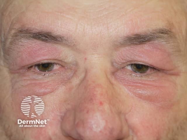
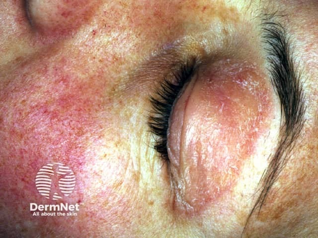
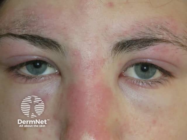
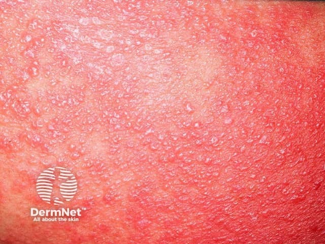
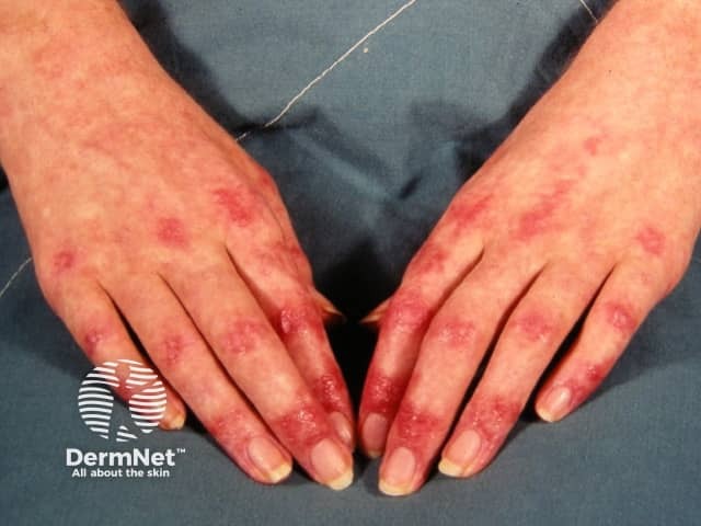
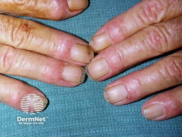
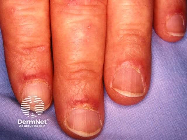
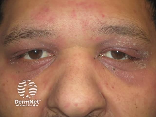
The skin changes of dermatomyositis will often, but not always, precede the muscle weakness. A face rash is the commonest initial skin sign, typically followed by scalp symptoms then the changes on the hands.
Cutaneous features of dermatomyositis
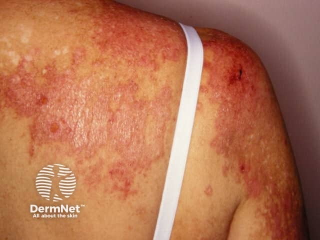
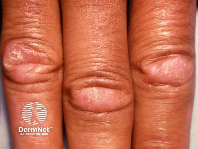
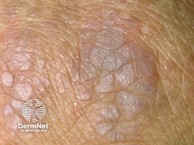
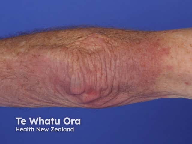
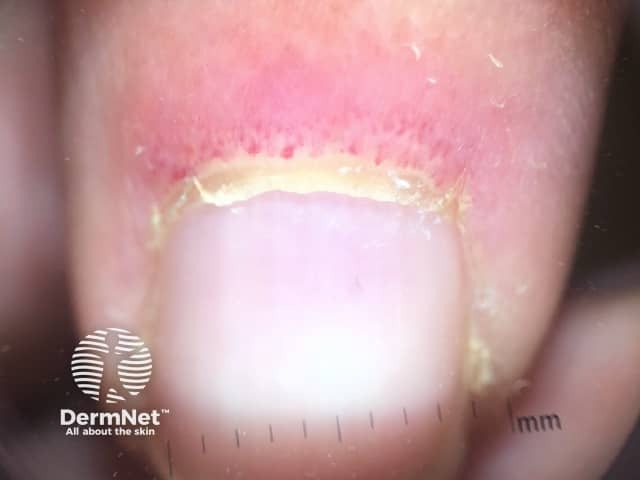
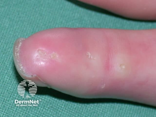
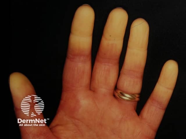
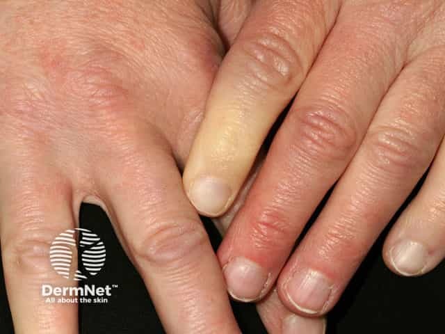
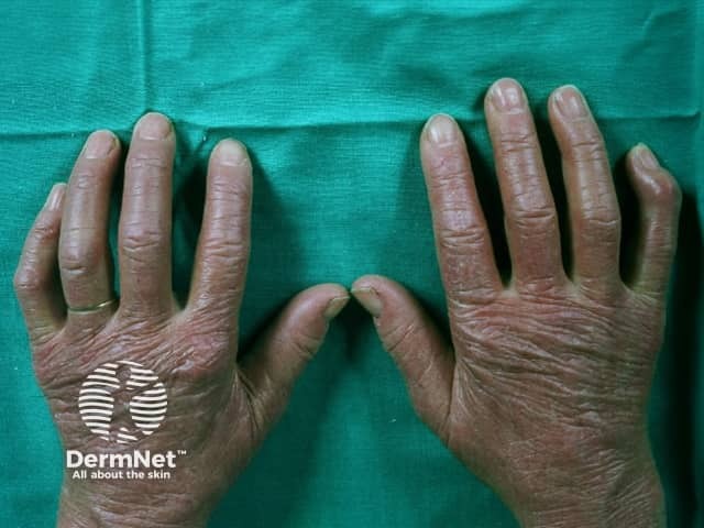
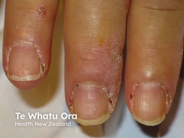
Characteristic skin changes of dermatomyositis include:
Heliotrope rash — bilateral lilac discolouration of the eyelids (particularly the upper eyelids) with swelling of the eyelids and skin around the eyes
Heliotrope rash
Heliotrope rash
Heliotrope rash
Shawl sign — fixed redness affecting the back, shoulders, chest, and neck which can become poikilodermatous
Shawl sign over the shoulder
Shawl sign over the upper back
Photosensitivity — sunburn reaction over the ‘V’ area of the anterior chest, face, and other sun-exposed sites
Photosensitive rash on the upper arm
Photosensitive rash on the V of chest
Gottron papules — purple papules and plaques over the tops of the small joints of the hands
Gottron papules over the joints of the hands
Gottron papules over finger joints
Violaceous hue of Gottron papules
Gottron sign — redness over the backs of the hands, elbows, and knees
Gottron sign over the knees
Gottron sign over the elbow
Gottron sign over a knee
Scalp involvement — itchy scaly scalp with inflammation and diffuse, non-scarring thinning of the hair, and sometimes poikiloderma
Scaly scalp in dermatomyositis
Scaly scalp with hair thinning in dermatomyositis
Nailfold changes — ragged cuticles and red nailfolds; dermoscopy demonstrates prominent blood vessels in the proximal nailfolds
Loss of cuticles with nailfold redness and Gottron papules
Ragged cuticles and nailfold redness
Nailfold dermoscopy showing dilated capillaries
Flagellate erythema
Holster sign – redness over the buttocks and lateral thighs
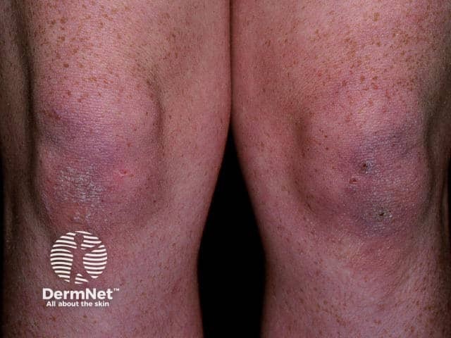
Muscle weakness may appear at the same time as the skin rashes, or it may occur weeks, months, or even years later. Amyopathic dermatomyositis presents with only skin changes and no clinical evidence of myositis, although this is usually found on investigation. The myositis usually affects the proximal muscles, those closest to the trunk as in the upper arms and thighs. The first indication of myositis is when the following every day movements become difficult with weakness and fatigue:
Climbing stairs or walking
Rising from a sitting or crouching position
Lifting objects
Raising arms above the shoulders, such as when combing hair
Difficulty swallowing.
The muscles may ache and become tender to touch.
How do clinical features vary in differing types of skin?
The heliotrope rash around the eyes characteristic of dermatomyositis can be difficult to distinguish in dark skin. Other clinical signs may be required to suggest the diagnosis.
Dermatomyositis of the hands
Heliotrope rash of the eyelids
Nailfold changes in dermatomyositis
What are the complications of adult-onset dermatomyositis?
When dermatomyositis affects swallowing, it may be complicated by:
Weight loss
Aspiration pneumonia.
Other systemic features of dermatomyositis may include:
Arthritis
Cardiac arrhythmias
Interstitial lung disease
Pulmonaryarterialhypertension.
How is adult-onset dermatomyositis diagnosed?
The diagnosis of dermatomyositis is suggested by the clinical features, and confirmed on investigations.
A skin biopsy of the rash shows an interfacedermatitis similar to cutaneous lupus erythematosus, so histology alone cannot be used to distinguish the two conditions. Dermoscopy or trichoscopy may also be used for closer inspection.
Blood tests assess the myositis and autoantibody subsets.
Anti-aminoacyl-transfer RNA synthetase antibodies — anti-Jo-1, anti-OJ, anti-EJ, anti-KS, anti-Zo, anti-Ha, anti-PL-1 are associated with antisynthetase syndrome.
Anti Mi-2 is associated with disease mainly affecting the skin.
Anti-transcriptional intermediary factor 1y antibodies (TIF-1y, anti 155/140) are strongly associated with malignancy-related dermatomyositis in the elderly.
Anti-melanocytedifferentiation-associated protein 5 (anti-MDA5) is associated with rapidly progressive interstitial lung disease and skin ulcers.
Anti-NXP2 (MJ) antibody is associated with pronounced muscle symptoms and less with skin changes.
Further investigation of the muscle disease may require:
Electromyography (EMG)
Muscle biopsy
Magnetic resonance imaging (MRI).
Age-appropriate investigations for an underlying malignancy should be considered after careful history and general examination. Low risk individuals should have basic blood and urine screening, and a plain chest X ray. Intermediate and high risk individuals should, in addition, receive a CT neck, thorax, abdomen and pelvis, pelvic and transvaginal ultrasound, cervical smear, mammography, prostate-specific antigen (PSA) test, cancer antigen 125 (CA-125) test and faecal occult blood test (FOBT)
What is the differential diagnosis for adult-onset dermatomyositis?
Dermatomyositis may need to be distinguished from:
Other idiopathic inflammatory myopathies, such as polymyositis or inclusion body myositis
The JAK inhibitor, baricitinib, was studied in a small, 12-patient, open-label trial with clinically significant improvement seen in as early as week 4 of the study. Apremilast was also studied in 8 adult women as add-on therapy with positive results.
Dazukibart, a monoclonal antibody targeting interferon β (IFNβ), demonstrated pronounced reduction of both muscle and skin disease in dermatomyositis in a phase II trial.
What is the outcome for adult-onset dermatomyositis?
Dermatomyositis may resolve in 20% of adults who have the condition, but most will require lifelong treatment. The prognosis of those with associated conditions such as malignancy, heart or lung involvement will be impacted and may be life-shortening.
Cobos GA, Femia A, Vleugels RA. Dermatomyositis: an update on diagnosis and treatment. Am J Clin Dermatol. 2020;21(3):339–53. doi:10.1007/s40257-020-00502-6. PubMed
DeWane ME, Waldman R, Lu J. Dermatomyositis: clinical features and pathogenesis. J Am Acad Dermatol. 2020;82(2):267–81. doi:10.1016/j.jaad.2019.06.1309. PubMed
Fiorentino, D., Mangold, A. R., Werth, V. P., Christopher-Stine, L., Femia, A., Chu, M., Musiek, A. C. M., Sluzevich, J. C., Graham, L. v, Fernandez, A. P., Aggarwal, R., Rieger, K., Page, K. M., Li, X., Hyde, C., Rath, N., Sloan, A., Oemar, B., Banerjee, A., … Vleugels, R. A. (2025). Efficacy, safety, and target engagement of dazukibart, an IFNβ specific monoclonal antibody, in adults with dermatomyositis: a multicentre, double-blind, randomised, placebo-controlled, phase 2 trial. The Lancet, 405(10473), 137–146. doi:10.1016/S0140-6736(24)02071-3. Journal
Okiyama N, Fujimoto M. Cutaneous manifestations of dermatomyositis characterized by myositis-specific autoantibodies. F1000Res. 2019;8:F1000 Faculty Rev-1951. doi:10.12688/f1000research.20646.1. PubMed Central
Papadakis MA, McPhee SJ, Rabow MW. Dermatomyositis. In: Current Medical Diagnosis and Treatment 2020, 59th edn, Lange, 2020: 864–7.
Sasaki H, Kohsaka H. Current diagnosis and treatment of polymyositis and dermatomyositis. Mod Rheumatol. 2018;28(6):913–21. doi:10.1080/14397595.2018.1467257. PubMed
Stone CJ, Faden DF, Xie L, et al. Application of Risk-Based Cancer Screening in Patients With Dermatomyositis. JAMA Dermatol. 2024;160(11):1248-1251. doi:10.1001/jamadermatol.2024.3355. PubMed
Thompson C, Piguet V, Choy E. The pathogenesis of dermatomyositis. Br J Dermatol. 2018;179(6):1256–62. doi:10.1111/bjd.15607. PubMed
Waldman R, DeWane ME, Lu J. Dermatomyositis: diagnosis and treatment. J Am Acad Dermatol. 2020;82(2):283–96. doi:10.1016/j.jaad.2019.05.105. PubMed
Zhao Q, Zhu Z, Fu Q, et al. Baricitinib for the treatment of cutaneous dermatomyositis: A prospective, open-label study. J Am Acad Dermatol. 2022;87(6):1374-1376. doi:10.1016/j.jaad.2022.08.025 Journal