
ADVERTISEMENT
You do not have any notes added to this page yet
Introduction and definitions
Benign disorders causing blisters and pustules in neonates
Viral infection
Bacterial infection
Fungal infection
Parasitic infection
Blistering from genodermatoses in neonates
Blistering from transient autoimmune diseases in neonates
Tests
Treatment
This page describes vesiculobullous and pustular lesions in newborns and their differentiating characteristics.
Vesiculobullous and pustular lesions in neonates can be due to miscellaneous benign conditions, an infection, a genodermatosis, or a transient autoimmune bullous disorder.
There are several benign disorders that may present within a few days of birth with blisters and pustules. These include:

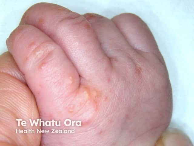
Neonatal blistering diseases may be due to viral, bacterial, fungal, or parasitic infection.
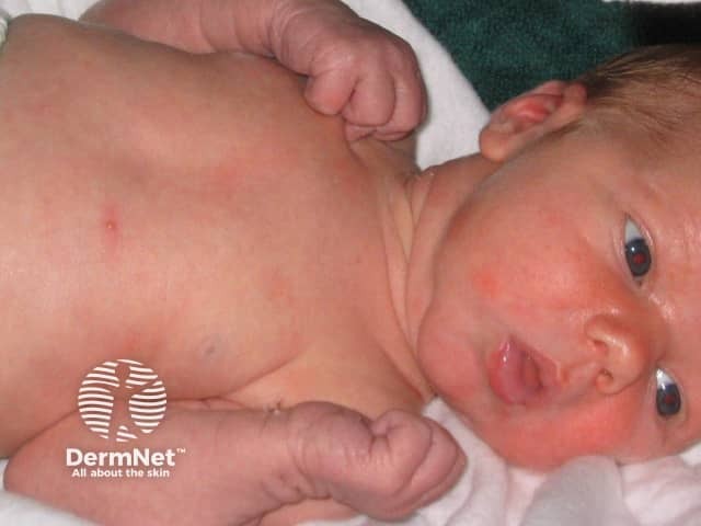
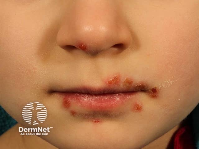
The onset of viral infections is within days to weeks after birth.
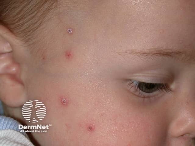
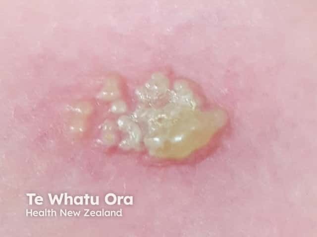
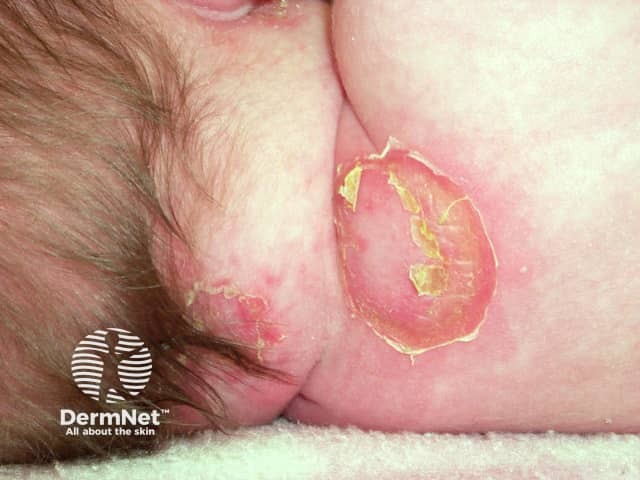
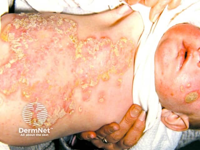
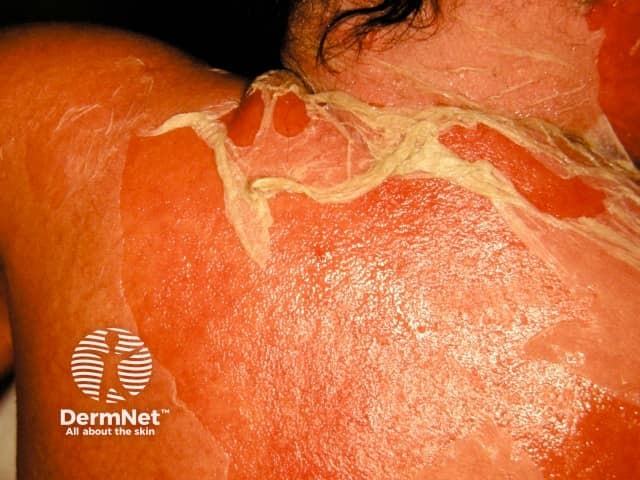
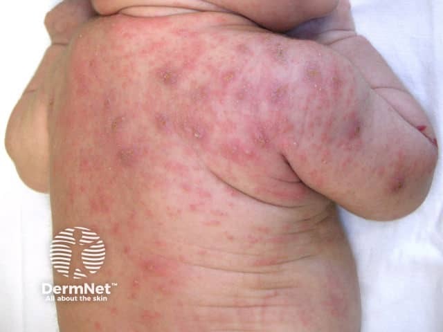
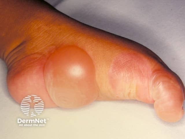
Staphylococcal infection in a neonate usually presents with localised superficial, flaccid, vesiculobullous or pustular lesions that rupture to reveal an erythematous base and then form seropurulent crusts. The infection may extend to cause fever and widespread SSSS [1].
Listeriosis is a cause of premature birth. It presents early with multiple pustules on the mucous membranes and skin and may progress to cause meningitis and septicaemia [1].
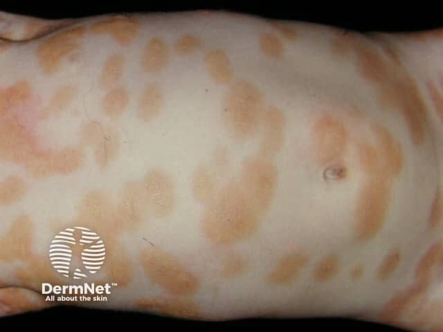
Congenital syphilis is associated with generalised haemorrhagic bullae and petechiae.
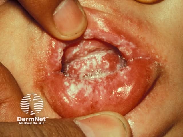
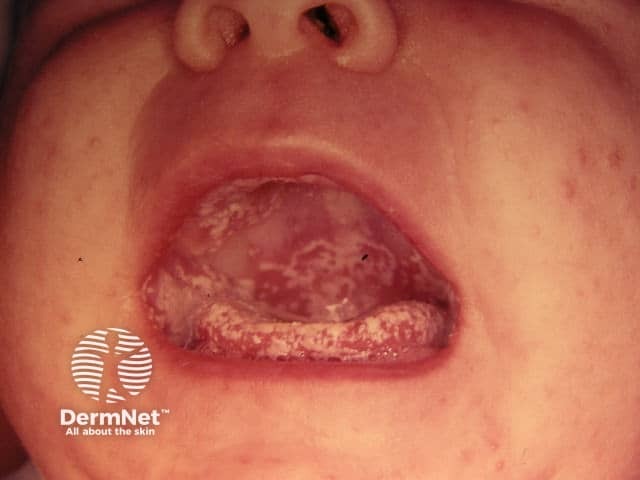
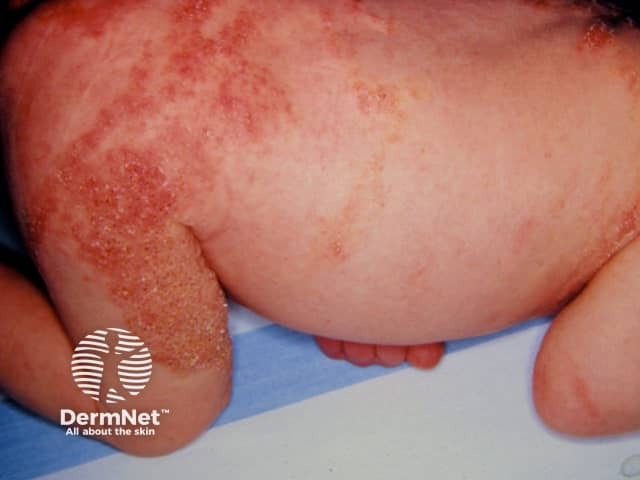
An infection caused by Candida albicans tends to occur a few weeks after birth or in an older baby, often presenting as oral thrush (white sticky plaques on a reddened mucosa) or napkin dermatitis. Candida infections are characterised by very superficial blisters and pustules associated with erythematous papules and plaques in intertriginous sites. Systemic mycosis with disseminated candida can also occur in neonates [1].
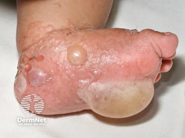
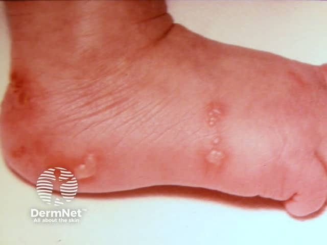
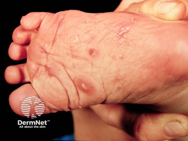
Scabies is caused by the parasitic mite Sarcoptes scabiei. In a young baby, it causes a widespread vesiculopustular eruption, that is prominent on the palms and soles. The source of the infestation is likely to be a family member or visitor with an itchy rash [1].
The blistering genodermatoses are:
Inherited vesiculopustular and bullous genodermatoses are rare. They should be suspected in newborns with a family history of a genodermatosis or consanguinity [2].
Maternal history of an autoimmune blistering disease can lead to a newborn presenting with the same autoimmune bullous disorder. Maternally transmitted autoimmune bullous disorders usually resolve within a few months of birth [1]. These include:
An initial investigation in a neonate with blisters includes scraping fluid and cells from an intact blister for viral/bacterial/fungal microscopy, culture, and testing for a polymerase chain reaction to specific organisms [1,2].
A skin biopsy, with or without direct immunofluorescence, should be undertaken if the infectious screen is negative and in those patients refractory to an initial therapy [3].
Blood, urine, and cerebrospinal fluid cultures can be used to detect disseminated disease in SSSS and herpes simplex [1].
Genodermatoses can be confirmed by skin biopsy using standard light microscopy, transmission electron microscopy, and immunofluorescence microscopy. Molecular genetic testing should also be considered [2].
An autoimmune blistering disease is investigated by a cord blood sample with serum indirect immunofluorescence on salt-split skin, and autoantibody enzyme-linked immunosorbent assay to desmoglein 1 and 3 and bullous pemphigoid antigen BP180. If there is no history of blistering in the mother, a lesional skin biopsy should be performed for histopathology. A perilesional skin biopsy should be submitted for direct immunofluorescence [2,4].
The treatment of the blistering disease depends on the diagnosis.
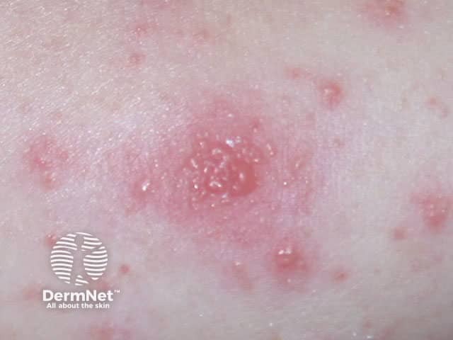
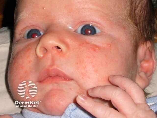
A benign disorder such as neonatal acne, erythema toxicum neonatorum, and transient neonatal pustular melanosis is self-limiting and no specific treatment is required [1].
Bacterial infection should be managed with empirical antibiotics according to each country's guidelines.
The management of epidermolysis bullosa is focused on prevention, wound care, and the optimisation of nutrition [2]. Aplasia cutis congenita is usually managed by conservative wound care.
Most cases of neonatal autoimmune blistering disease are self-limiting, and symptoms may be reduced by the use of topical corticosteroids [3].
An AI summary will appear based on your search term using data from all of the topic pages across the entire DermNet site.
Show more