Skin, hair and nail tissue are collected for microscopy and culture (mycology) to establish or confirm the diagnosis of a fungal infection.
Exposing the site to long-wavelength ultraviolet radiation (Wood lamp) can help identify some fungal infections of hair (tinea capitis) because the infected hair fluoresces green.
Specimen collection for fungal testing
Specimens for fungal microscopy and culture are transported to the laboratory in a sterile container or a black paper envelope. They include:
Scrapings of scale, best taken from the leading edge of the rash after the skin has been cleaned with alcohol
Skin stripped off with adhesive tape, which is then stuck on a glass slide
Hair which has been pulled out from the roots
Brushings from an area of scaling in the scalp
Nail clippings or skin scraped from under a nail
A skin biopsy
A moist swab from a mucosal surface (inside the mouth or vagina) in a special transport medium.
A swab should also be taken from pustules in case of secondary bacterial infection.
Direct microscopy of skin scrapings and nail clippings
The material is examined by microscopy by one or more of these methods:
Potassium hydroxide (KOH) preparation, stained with blue or black ink
Fluorescent staining
An unstained wet-mount
A stained dried smear
Histopathology of biopsy with special stains, such as periodic acid-Schiff (PAS).
Microscopy can identify a dermatophyte by the presence of:
Fungal hyphae (branched filaments) making up a mycelium
Arthrospores (broken-off spores)
Arthroconidia (specialised external spores)
Spores inside a hair (endothrix) or outside a hair (ectothrix).
Fungal elements are sometimes difficult to find, especially if the tissue is very inflamed, so a negative result does not rule out fungal infection.
A yeast infection can be identified by the presence of:
Yeast cells, which may be dividing by budding
Pseudohyphae (branched filaments similar to those of a dermatophyte) forming a pseudomycelium.
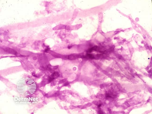
PAS stain of aspergillus seen in a skin biopsy
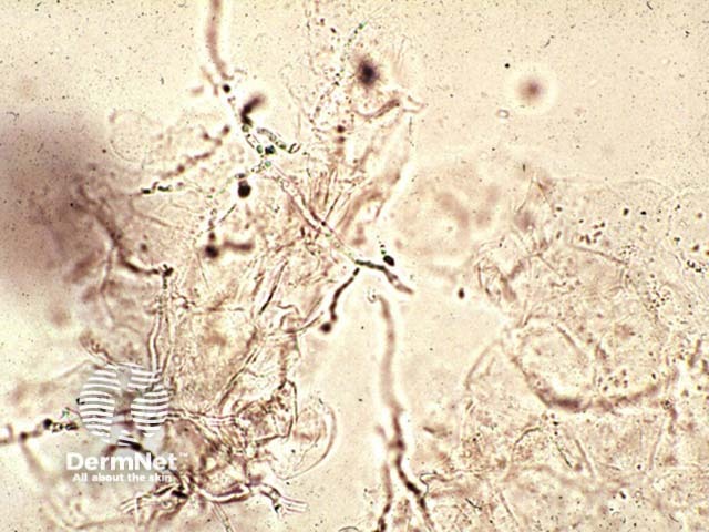
KOH of M. canis
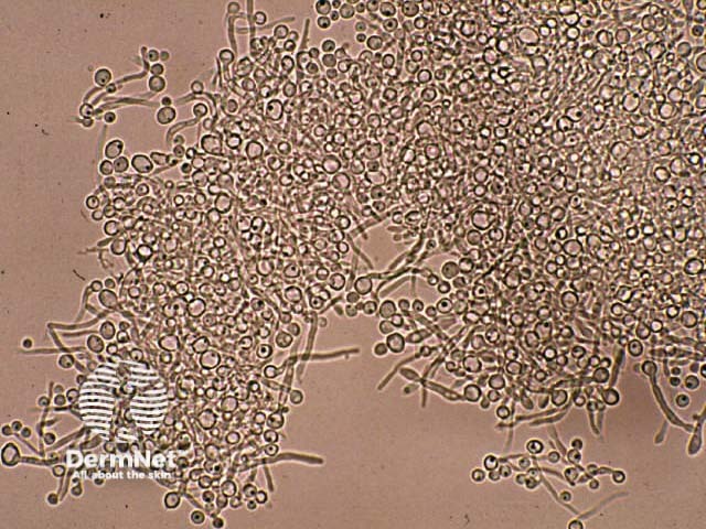
KOH of candida
Culture of fungi
Culture identifies which organism is responsible for the infection:
To find out the source of infection e.g. a particular animal
To select the most suitable treatment.
Growing the fungus in culture may take several weeks, incubated at 25–30ºC. The specimen is inoculated into a medium such as Sabouraud's dextrose agar containing cycloheximide and chloramphenicol. The cycloheximide is left out if a mould requires identification.
A negative culture may arise because:
The condition is not due to fungal infection.
The specimen was not collected properly.
Antifungal treatment had been used before the collection of the specimen.
There was a delay before the specimen reached the laboratory.
The laboratory procedures were incorrect.
The organism grows very slowly.
The culture of yeasts and moulds may be due to harmless colonisation rather than infection; this is common in an underlying skin disease such as psoriasis.
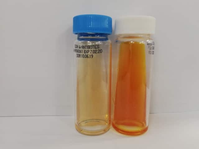
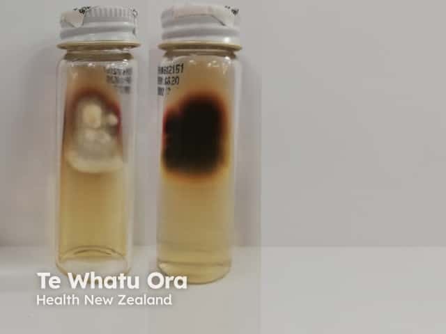
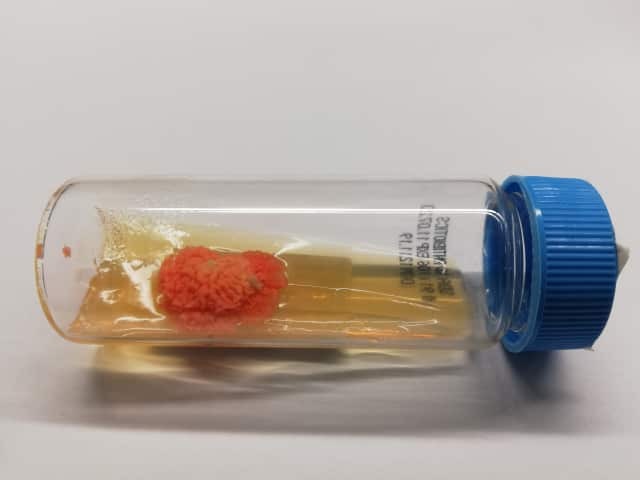
Culture slopes
Trichophyton rubrum culture on agar slope
Blood tests for patients with deep or disseminated fungal infection
Blood tests are not useful for the diagnosis of superficial fungal infections. In subcutaneous and systemic mycoses, several tests may be helpful.
Molecular testing by the PCR technique has become widespread in recent years and has a higher sensitivity than either culture or direct microscopy.
Results are often available in 24 hrs, and give fungal species identification that it useful both therapeutically and epidemiologically.
Bibliography
Paugam A, L'ollivier C, Viguié C, et al. Comparison of real-time PCR with conventional methods to detect dermatophytes in samples from patients with suspected dermatophytosis. J Microbiol Methods. 2013;95(2):218-222. Journal