Author: Dr Ahmed Sadek, Dermatologist, Cairo Hospital for Dermatology & Venereology (Al-Haud Al-Marsoud), Egypt (2022)
Contributors: Dr Dalia Hossam, Dr Radwa Magdy, Dr Nehal Saied, Dr Noha Hashem, Dr Safaa Yehia Negm, Dr Moshera Saied El Bahrawy, Dr Amira Ragab, Dr Amal Wagih, Dr Haidy El-Hussieny, Dr Mona Ragib, Dr Hala Amer (2022)
This page covers the diagnosis of different structural genetichair shaft defects. These are classified based on their association with hair fragility.
Structural hair shaft defects associated with hair fragility
Monilethrix
The term is derived from the Greek meaning “necklace” or “beaded”. Monilethrix is an autosomal dominant disorder that usually affects individuals in the first few months after birth. Affected hairs appear short, fragile, and brittle. The disorder mainly affects the scalp, however, in severe cases it may involve eyebrows, eyelashes, and nails (koilonychia).
Trichoscopic examination
Affected hairs show abnormal constrictions regularly arranged along the hair shaft causing variability in the shaft thickness and its breakage at the constriction sites
Medulla is present in nodes and absent in internode segments
Regrowing hair (pigtail hair)
Perifollicular casts
Horny follicularpapules appear as large yellow dots when examined with immersion fluid
Follicular plugging, perifollicular erythema and scales observed when examined using dry dermoscopy.
Monilethrix presenting with constrictions regularly arranged along the hair shaft (DHS-patient1)
Monilethrix-like hairs
Monilethrix-like hairs show the same constrictions but without the regular distribution characteristic of true monilethrix. It occurs in monilethrix-like congenitalhypotrichosis, alopecia areata, lichen planopilaris and patients receiving chemotherapy.
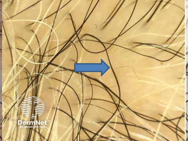
Trichorrhexis invaginata (bamboo hair)
An abnormality of the hair in which the hair shaft invaginates into itself at multiple points along the shaft. The disorder becomes evident at infancy. Affected hairs are short, sparse, and very fragile. It is pathognomonic for Netherton syndrome. Recently, matchstick hairs were also described in Netherton syndrome as short hairs with a bulging end.
Trichoscopic examination
Multiple small nodules along the hair shaft observed with handheld dermoscopy.
Using higher magnification, affected hair show invagination of the distal portion of the shaft into its proximal portion, forming a “ball-in-cup” appearance
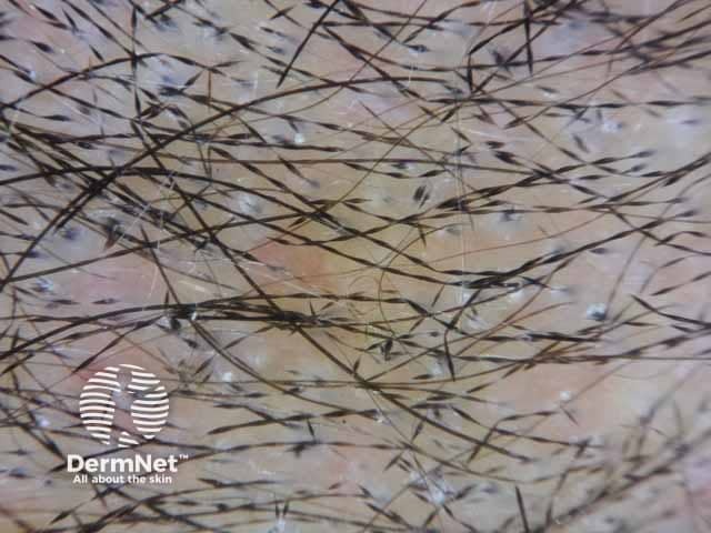
Trichorrhexis nodosa
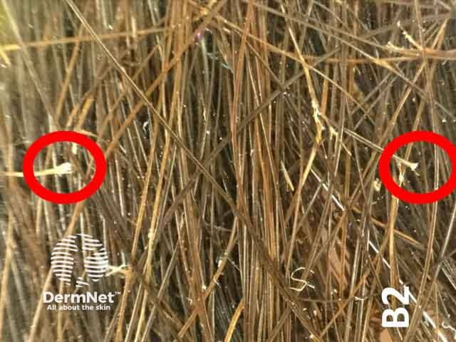
The most common structural hair shaft abnormality caused by damage of the hair shaft cuticle. Causes:
Tricho-hepato-enteric syndrome
Arginino-succinic-aciduria
Ectodermal dysplasias
Monilethrix-like congenital hypotrichosis
Mutation in the XPD gene
Congenital trichorrhexis nodosa without coexisting defects.
Trichoscopic examination
The trichoscopic features of trichorrhexis nodosa depend on the magnification used and the presence of immersion fluid (Fig 50).
Using a handheld dermatoscope, affected hairs show light colored nodular thickenings along the hair shafts.
Higher magnification devices show splitting of hair shafts into multiple fibers appearing like two brushes pushed into one another.
When a hair shaft breaks at the site of the nodule, it leaves a slightly thickened, rounded hair shaft end, which may appear darker compared with light-colored hair shaft.
At higher magnification, trichoscopy allows appreciating numerous brush-like small fibers at the distal end of the hair shaft.
Brush-like small fibers at the distal end of the hair shaft (red circles) denoting trichorrhexis nodosa in a female patient (DHS-patient2)
Pili torti
Pili torti is a structural hair shaft defect which is associated with multiple disorders.
Conditions associated with pili torti:
Pili torti
Ronchese type
Beare type
Autosomal recessiveichthyosis with hypotrichosis (ARIH)
Basex syndrome
Björnstad syndrome
Crandall syndrome
Hypohidrotic ectodermal dysplasia
Menkes syndrome
Hypotrichosis with juvenilemaculardystrophy
Laron syndrome.
Trichoscopic examination
Affected hair shafts show flattening with twisting of the hair fiber three to ten twists on its long axis. This abnormality is best observed by dry dermoscopy at high magnification.
Pili torti; flattened and twisted hairs on their own axis
Trichothiodystrophy
It is an autosomal recessive disorder characterised by sulfur-deficient hair. This disorder is mainly diagnosed using polarized light microscopy as it shows alternating bright and dark bands along the hair shaft, resembling a tiger’s tail.
Trichoscopic examination
Trichoscopic examination is of limited value in diagnosing the disorder; it shows heterogeneous hair shafts resembling grains of sand with slightly wavy contour.
Structural hair abnormalities not associated with hair fragility
Pili annulati
Pili annulati is characterized by multiple air-filled cavities along the hair shaft. The disorder is detected in blonde hair much more easily than in darkly pigmented hair.
Trichoscopic examination
Alternating dark and light bands along the hair shaft corresponding to the air-filled cavities.
Woolly hair
Hutchinson et al. classified the condition into three variants:
This disorder is characterised by tightly curled hair shafts with 180° longitudinal twisting, giving the appearance of a crawling snake. Common associations:
Ankad BS, Mukherjee SS. Hair Shaft Defect in a Teenage Girl: Trichoscopy Saves the Day!. Dermatol Pract Concept. 2020;11(1):e2020080. Published 2020 Dec 7. doi:10.5826/dpc.1101a80. Journal
Boussofara L, Ghannouchi N, et al. Netherton's syndrome: the importance of eyebrow hair. Dermatol Online J. 2007;13(3):21. Published 2007 Jul 13. Abstract
Erez A, Nagamani SC, Lee B. Argininosuccinate lyase deficiency-argininosuccinic aciduria and beyond. Am J Med Genet C Semin Med Genet. 2011;157C(1):45–53. doi:10.1002/ajmg.c.30289. Journal
Fabre A, André N, et al. Intractable diarrhea with "phenotypic anomalies" and tricho-hepato-enteric syndrome: two names for the same disorder. Am J Med Genet A. 2007;143A(6):584–8. doi:10.1002/ajmg.a.31634. Journal
Hays SB, Camisa C. Acquired pili torti in two patients treated with synthetic retinoids. Cutis. 1985;35(5):466–8. Abstract
Itin PH, Sarasin A, Pittelkow MR. Trichothiodystrophy: update on the sulfur-deficient brittle hair syndromes. J Am Acad Dermatol. 2001;44(6):891-924. doi:10.1067/mjd.2001.114294. Abstract
Mirmirani P. Ceramic flat irons: improper use leading to acquired trichorrhexis nodosa. J Am Acad Dermatol. 2010;62(1):145–7. doi:10.1016/j.jaad.2009.01.048. Journal
Neila Iglesias J, Rodríguez Pichardo A et al. Masquerading of trichotillomania in a family with monilethrix. Eur J Dermatol. 2011;21(1):133. doi:10.1684/ejd.2010.1194. Journal
Rakowska A, Kowalska-Oledzka E et al. Hair shaft videodermoscopy in netherton syndrome. Pediatr Dermatol. 2009;26(3):320–2. doi:10.1111/j.1525-1470.2008.00778.x. Journal
Rakowska A, Slowinska M, et al. Dermoscopy as a tool for rapid diagnosis of monilethrix. J Drugs Dermatol. 2007;6(2):222–4. Abstract
Rakowska A, Slowinska M, et al. Trichoscopy in genetic hair shaft abnormalities. J Dermatol Case Rep. 2008;2(2):14–20. doi:10.3315/jdcr.2008.1009. Journal
Rouse C, Siegfried E, et al. Hair and sweat glands in families with hypohidrotic ectodermal dysplasia: further characterization. Arch Dermatol. 2004;140(7):850–5. doi:10.1001/archderm.140.7.850. Journal
Rudnicka L, Olszewska M, Waśkiel A, Rakowska A. Trichoscopy in Hair Shaft Disorders. Dermatol Clin. 2018;36(4):421–30. doi:10.1016/j.det.2018.05.009. Journal
Stratigos AJ, Baden HP. Unraveling the molecular mechanisms of hair and nail genodermatoses. Arch Dermatol. 2001;137(11):1465–71. doi:10.1001/archderm.137.11.1465. Journal
Whiting DA, Dy LC. Office diagnosis of hair shaft defects. Semin Cutan Med Surg. 2006;25(1):24-34. doi:10.1016/j.sder.2006.01.004. PubMed
Zitelli JA. Pseudomonilethrix. An artifact. Arch Dermatol. 1986;122(6):688-690. doi:10.1001/archderm.122.6.688. Journal