
ADVERTISEMENT
You do not have any notes added to this page yet
Introduction
Demographics
Causes
Clinical features
Variation in skin types
Complications
Diagnosis
Differential diagnoses
Treatment
Prevention
Outcome
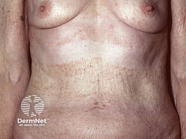
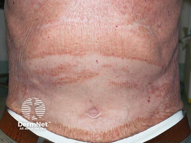
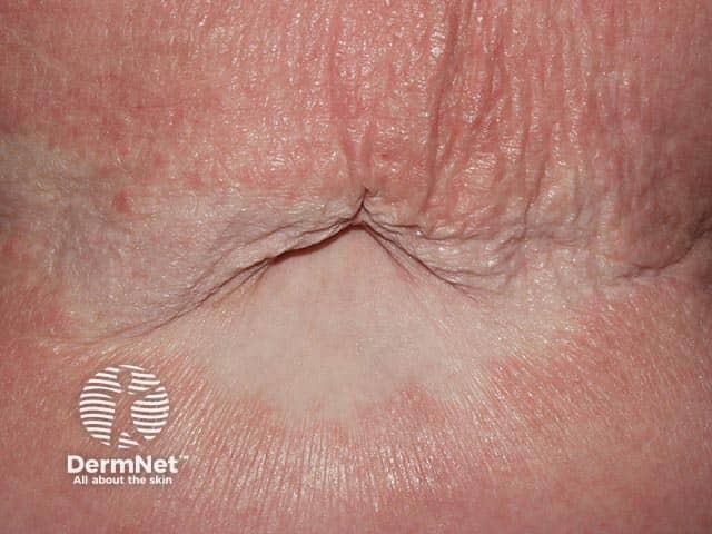
Papuloerythroderma of Ofuji (PEO) is a rare inflammatory skin disorder characterised by widespread pruritic papular eruptions that coalesce into larger plaques sparing the skin folds. This produces the “deck-chair sign”, which is pathognomonic of PEO.
Click here for more images of papuloerythroderma of Ofuji
Papuloerythroderma of Ofuji is reported to affect approximately 1.5 individuals per million. However, as this condition is poorly understood, this number is likely an underestimate.
PEO is also:
PEO is commonly found in individuals with underlying lymphomas such as cutaneous T-cell lymphoma (CTCL). Other associations include solid organ malignancies, underlying atopy, drugs, and infections (HIV, hepatitis C).
The cause of papuloerythroderma of Ofuji is yet to be fully understood. When no underlying cause can be identified, it is termed ‘Idiopathic’ or ‘primary’. Of all published cases, approximately half are of primary aetiology (ie,. idiopathic) and the remaining half is secondary PEO. Secondary PEO incorporates atopy, infection, malignancy (ie,. gastric carcinoma, hepatocellular carcinoma, adenocarcinoma of the colon, prostate cancer and chronic lymphocytic leukaemia) and drug-induced PEO.
The strong association between PEO and CTCL leads to the possibility of PEO being a paraneoplastic syndrome, therefore screening for underlying malignancies is important during patient work-up. Immune system dysregulation, with a specific role played by T-Helper (Th) 2 and Th22, appears to be important in the onset of PEO.
Hepatitis C and HIV have also been found to cause PEO with complete resolution of all lesions after levels of the viruses became undetectable. Drug-induced papuloerythroderma is attributed to Th2 cell-mediated hypersensitivity and has been linked to medications such as furosemide, aspirin, diltiazem, didanosine, nicardipine, isoniazid, ranitidine, leuprorelin, ticlopidine, eperisone, and etretinate. Likewise, immune dysregulation and the involvement of the Th2/Th22 axis are considered contributing factors in the association between atopy and PEO, with nearly 20% of PEO patients displaying atopy.
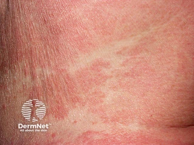
Papuloerythroderma of Ofuji presents as a widespread pruritic eruption involving the trunk, extremities, and head. Polygonal, flat papules with hues ranging from pink to brown may coalesce, evolving into erythroderma in a cobblestone pattern. Sparing of the face and skin folds produces the 'deck-chair sign', considered typical of PEO.
Other clinical features include:
Erythema tends to be less discernible in individuals with darker skin types. In individuals with Fitzpatrick skin types IV, V, and VI, erythematous areas tend to present as a more violaceous hue or a darker brown, in contrast to individuals with Fitzpatrick skin types I to III, where the tones are towards pink or red. Consequently, visible signs of erythema or inflammation may be absent, with manifestations limited to excoriation and lichenification.
Complications of papuloerythroderma of Ofuji include secondary bacterial infections from excoriation and delay of diagnosis of CTCL.
Primary papuloerythroderma of Ofuji is a diagnosis of exclusion. Controversy exists as to whether PEO is an independent entity or a clinical manifestation seen in different conditions. Notably, skin lesions often persist for over a year before a diagnosis is established.
A diagnostic criterion published by Torchia et al. remains a key component to the diagnosis of primary papuloerythroderma of Ofuji. The components of the criteria include:
Other features frequently observed in PEO include age over 55 years, male gender, tissue and/or peripheral eosinophilia, elevated immunoglobulin E (IgE), and peripheral lymphopenia.
Dermoscopy of PEO may show several erythematous pinpoint papules surrounded by white halos against a erythematous-pinkish background.
Histology is non-specific and may reveal a spongiotic reaction pattern, along with a dense dermal perivascular infiltrate consisting of lymphocytes, histiocytes and eosinophils. Inflammatory cell exocytosis may be present, however, it is essential to differentiate this from the epidermotropism observed in CTCL. To rule out CTCL, histopathologic examination alone may not always be diagnostic, and in such cases, additional studies such as flow cytometry and T-cell receptor clonality studies are essential.
Treat underlying causes that are identified and stop offending drugs.
PEO can be, but is infrequently a serious condition necessitating hospital admission for close monitoring. General measures including frequent vital sign monitoring, fluid and electrolyte balance assessments, maintenance of skin hydration using wet wraps and emollients, and simple analgesia in those where the erythroderma is producing systemic effects .
Currently, there is no established treatment guideline for idiopathic PEO. However, various therapeutic approaches have demonstrated positive outcomes, including:
Notably, combination therapies have proven to be more effective than single interventions, with the combination of PUVA with either topical corticosteroids or oral retinoids displaying the highest complete resolution rate. Recent cases have also reported successful treatment of PEO with dupilumab after exclusion of underlying CTCL.
In most cases, papuloerythroderma of Ofuji cannot be prevented.
Patients with known offending medication should avoid the drug forever and medical records should be updated. Patients with infections, malignancies or atopy associated with PEO should be informed if they are at known risk.
Idiopathic papuloerythroderma of Ofuji typically leads to remission a few years after onset, with a mean duration of 2.6 years. Secondary PEO may lead to a faster complete resolution upon addressing the underlying causes.
All patients with PEO should undergo screening to rule out malignancies and infections.
An AI summary will appear based on your search term using data from all of the topic pages across the entire DermNet site.
Show more