Authors: Dr Amanda Oakley MBChB FRACP, Dept of Dermatology Waikato Hospital, New Zealand (2004); Dr Tyler Stevens, Counties Manukau DHB, New Zealand (2023).
Dermoscopy or dermatoscopy refers to the examination of the skin using skin surface microscopy, and is also called ‘epiluminoscopy’ and ‘epiluminescent microscopy’. Derm(at)oscopy is mainly used to evaluate pigmented skin lesions. In experienced hands it can make it easier to diagnose melanoma.
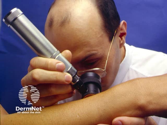
Dermoscopy requires a high quality magnifying lens and a powerful lighting system (a dermatoscope). See below for more information on dermatoscopes.
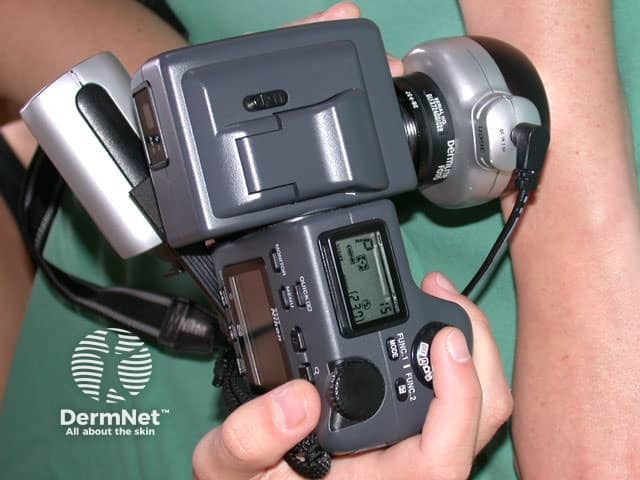
Computer software can be used to archive dermoscopy images and allow expert diagnosis and reporting (mole mapping). Smart programs may aid in diagnosis by comparing the new image with stored cases with typical features of benign and malignantpigmented skin lesions.
Skin examination
Photography
Dermatoscopic features of pigmented lesions
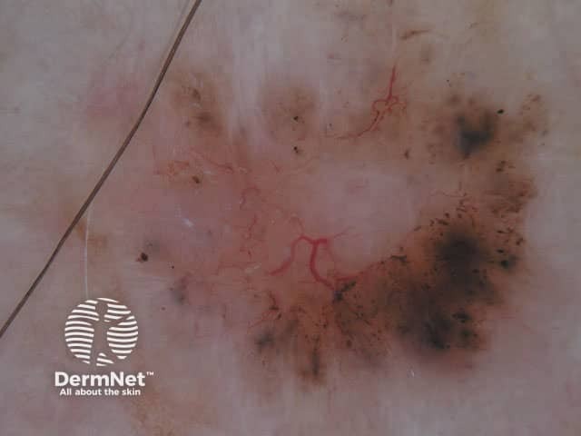
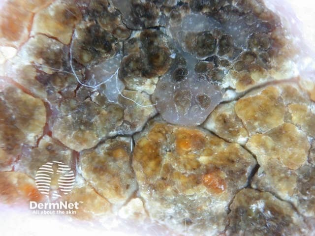
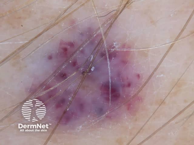
Using dermoscopy, the pigmentation of the lesion is evaluated in terms of colour(s) and structure.
Colours found in pigmented skin lesions include black, brown, red, blue, grey, yellow and white.
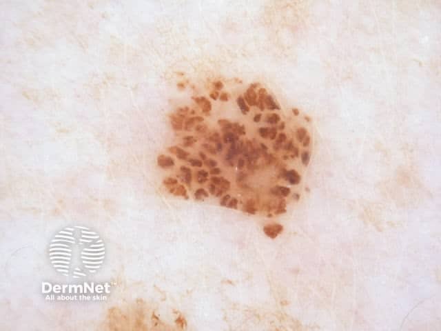
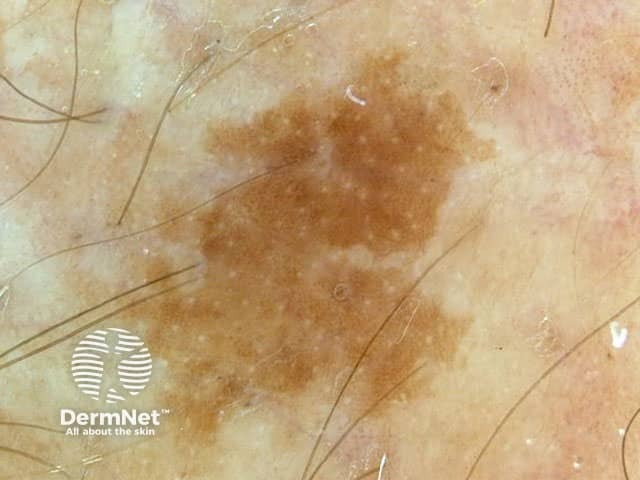
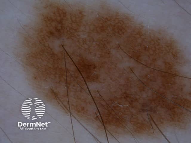
Common naevus
Characteristics of the dermatoscopic structure of the skin lesions include:
Symmetry or asymmetry
Homogeny/uniformity (sameness) or heterogeny (structural differences across the lesion)
Distribution of pigment: brown lines, dots, clods and structureless areas
Skin surface keratin: small white cysts, crypts, fissures
Vascularmorphology and pattern: regular or irregular
Border of the lesion: fading, sharply cut off or radial streaks
Presence of ulceration
There are specific dermoscopic patterns that aid in the diagnosis of the following pigmented skin lesions:
Used widely by dermatologists, plastic surgeons, and general practitioners, dermatoscopes allow the user to perform skin surface microscopy. This enables the practitioner to examine skin structures and patterns.
There are several different dermatoscopes and these are either conventional standalone devices, or smartphone attachable. When choosing a dermatoscope, there are some important things to consider:
Compatibility - smartphones and cameras
Polarisation - polarised, linear-polarised, unpolarised, or variable
Contact or Non-contact viewing - or both
Magnification - eg, 10x, 16x, or 40x
Battery - disposable, rechargeable, time to charge, time of continuous operation
Use dermoscopy to examine all pigmented lesions referred for assessment and ensure that all staff are adequately trained in its use. Melanoma: summary of NICE guidance