Author: Dr Fiona Larsen, Dermatology Registrar, Green Lane Hospital, Auckland, New Zealand, 2005. Updated by Dr Janice Yeon, Dermatology Research Fellow, The Skin Hospital, Sydney NSW, Australia. DermNet Editor in Chief: Adjunct A/Prof Amanda Oakley, Dermatologist, Hamilton, New Zealand. Copy edited by Gus Mitchell. October 2020.
What are the clinical features of eosinophilic cellulitis?
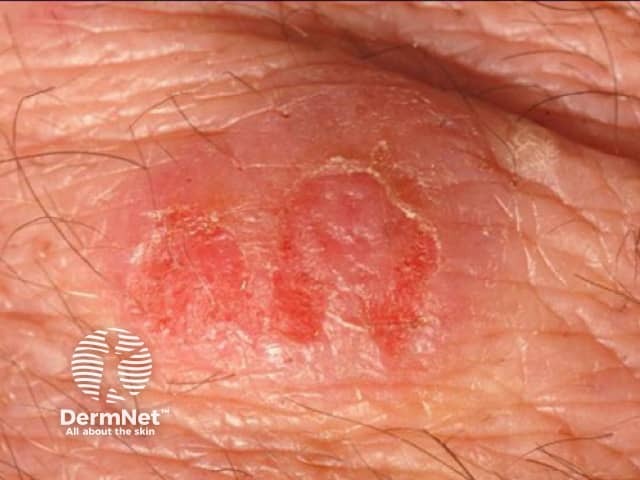
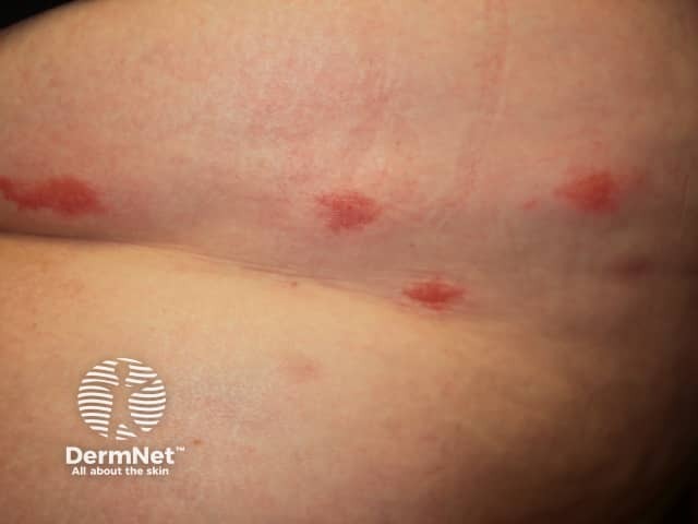
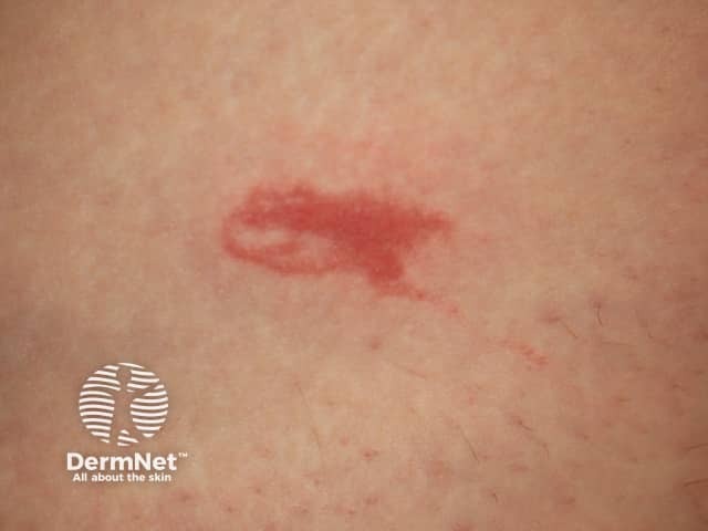
Eosinophilic cellulitis usually presents as itchy or burning erythematousnodules or plaques resembling cellulitis or erysipelas on a limb. However, the clinical appearance is variable: lesions can be single or multiple, the face and trunk may be affected, and clinical morphology can include:
Plaque type
Annulargranuloma-like
Urticaria-like
Papulovesicular
Bullous
Papulonodular
Fixed drug eruption-like.
The classic plaque-type variant of eosinophilic cellulitis is the most common presentation in children, whereas the annular granuloma-like variant is more frequently seen in adults.
Individual lesions can resolve spontaneously but typically recur.
Associated systemic symptoms may include fever and arthralgias.
What are the complications of eosinophilic cellulitis?
Eosinophilic cellulitis may be rarely complicated by superinfection.
Severe swelling of a limb can cause compartment syndrome.
How is eosinophilic cellulitis diagnosed?
Eosinophilic cellulitis is often misdiagnosed initially as cellulitis or erysipelas, and is only considered when antibiotic treatment is unhelpful.
Proposed diagnostic criteria for eosinophilic cellulitis require at least two major and one minor criteria.
Major criteria
Major criteria for an eosinophilic cellulitis diagnosis include:
Clinical picture to include any of the reported variants
No evidence of systemic disease
Relapsing, remitting course
Histology comprises eosinophilic infiltrates without vasculitis.
Minor criteria
Minor criteria for an eosinophilic cellulitis diagnosis include:
Peripheral eosinophilia not persistent and not greater than >1.5 x 10⁹/L
Histology has granulomatous change
Flame figures
A triggering factor (eg, a drug).
Peripheraleosinophilia affects approximately 50% of cases but is not required for diagnosis.
Skin biopsy is usually required and the histological findings depend on the stage of disease.
Key findings in the acute phase are dermaloedema with an eosinophilic infiltrate and without vasculitis.
The subacute phase shows the characteristic ‘flame figures’.
The chronic phase is granulomatous with histiocytes and giant cells (see Wells syndrome pathology).
Flame figures are not pathognomonic of Wells syndrome and can be seen with other eosinophilic infiltrates, such as:
Treatment of an associated trigger, such as cessation of an implicated drug, can lead to complete resolution.
What is the outcome for eosinophilic cellulitis?
Eosinophilic cellulitis has a benign course with a tendency for lesions to spontaneously resolve. However, recurrence is common.
References
Wells GC, Smith NP. Eosinophilic cellulitis. Br J Dermatol. 1979;100(1):101–9. doi:10.1111/j.1365-2133.1979.tb03574.x. PubMed
Heelan K, Ryan JF, Shear NH, Egan CA. Wells syndrome (eosinophilic cellulitis): proposed diagnostic criteria and a literature review of the drug-induced variant. J Dermatol Case Rep. 2013;7(4):113–20. doi:10.3315/jdcr.2013.1157. PubMed Central
Nielsen T, Schmidt H, Søgaard H. Eosinophilic cellulitis. (Well's syndrome) in a child. Arch Dermatol. 1981;117(7):427–9. Journal
Caputo R, Marzano AV, Vezzoli P, Lunardon L. Wells syndrome in adults and children: a report of 19 cases. Arch Dermatol. 2006;142(9):1157–61. doi:10.1001/archderm.142.9.1157. PubMed
Anderson CR, Jenkins D, Tron V, Prendiville JS. Wells' syndrome in childhood: case report and review of the literature. J Am Acad Dermatol. 1995;33(5 Pt 2):857–64. doi:10.1016/0190-9622(95)90423-9. Journal
Fujii K, Tanabe H, Kanno Y, Konishi K, Ohgou N. Eosinophilic cellulitis as a cutaneous manifestation of idiopathic hypereosinophilic syndrome. J Am Acad Dermatol. 2003;49(6):1174–7. doi:10.1016/s0190-9622(03)00466-3. PubMed
Räßler F, Lukács J, Elsner P. Treatment of eosinophilic cellulitis (Wells syndrome) - a systematic review. J Eur Acad Dermatol Venereol. 2016;30(9):1465–79. doi:10.1111/jdv.13706. PubMed
Yeon J, Chan RC, Zagarella S. Eosinophilic cellulitis successfully treated with methotrexate [published online ahead of print, 2020 Jun 25]. Australas J Dermatol. 2020;10.1111/ajd.13358. doi:10.1111/ajd.13358. Journal
Sinno H, Lacroix JP, Lee J, et al. Diagnosis and management of eosinophilic cellulitis (Wells' syndrome): a case series and literature review. Can J Plast Surg. 2012;20(2):91–7. doi:10.1177/229255031202000204. PubMed Central