
ADVERTISEMENT
You do not have any notes added to this page yet
Abnormalities of the nail plate surface Nail discolouration Abnormalities of the cuticle and nail fold Abnormalities of nail shape Loss of nails Lesions around nails
This page outlines the terms used by dermatologists to describe diseases of the fingernails and toenails.
Nail plate abnormalities are often due to inflammatory conditions affecting the matrix or nail bed. Specific diagnoses may be made from characteristic appearances.
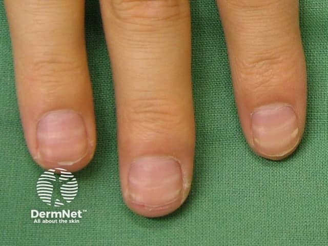
Nail pitting may be a sign of eczema, psoriasis and alopecia areata.
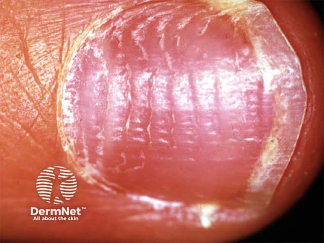
Transverse ridging of a nail may be a sign of eczema, paronychia, psoriasis, parakeratosis pustulosa, and shrimp nail.
A Beau line is a transverse depression affecting all nails, due to acute systemic illness stopping nail growth.
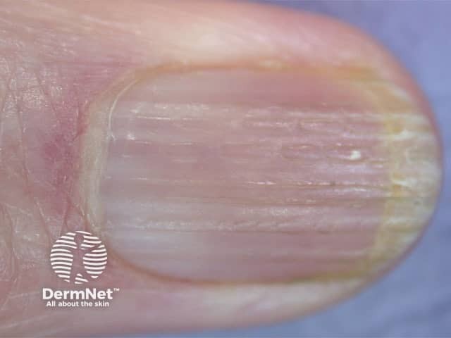
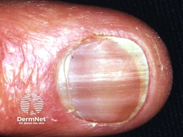
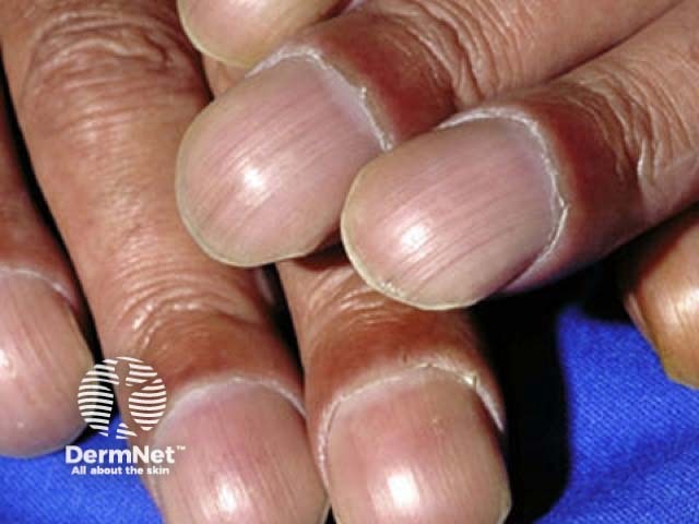
Onychorrhexis is longitudinal ridging of the nail plate. Consider ageing, lichen planus, psoriasis, fungal nail infection and Darier disease or a habit of picking.
A horizontal groove can be the result of earlier nail trauma or subungual haematoma.
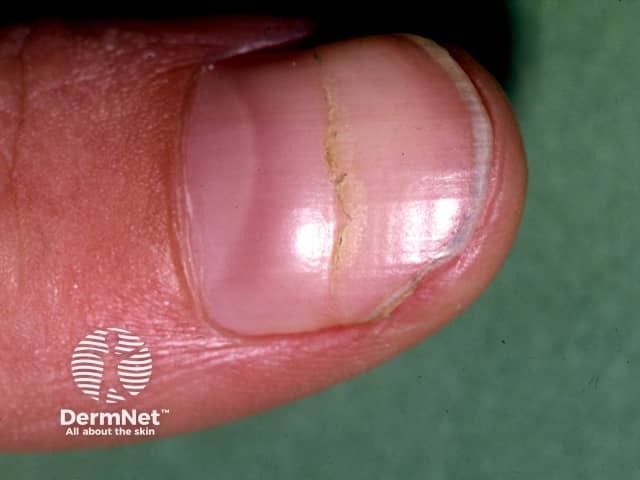
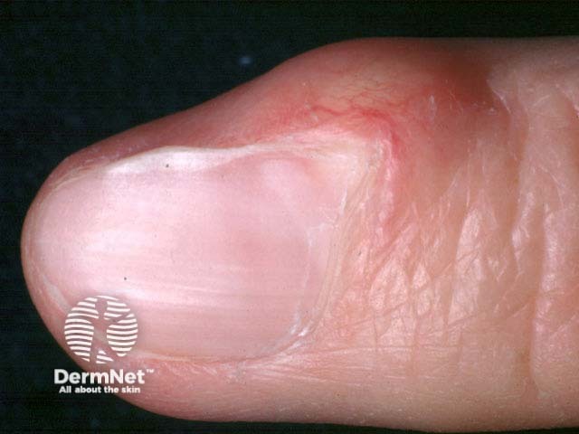
A longitudinal groove in the nail plate is due to myxoid cyst or wart overlying the proximal nail matrix.
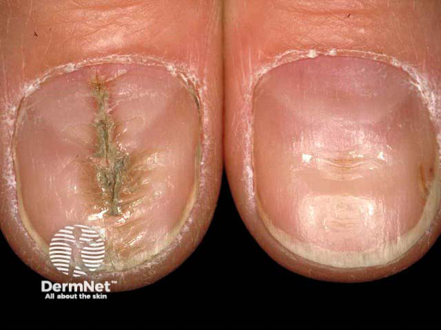
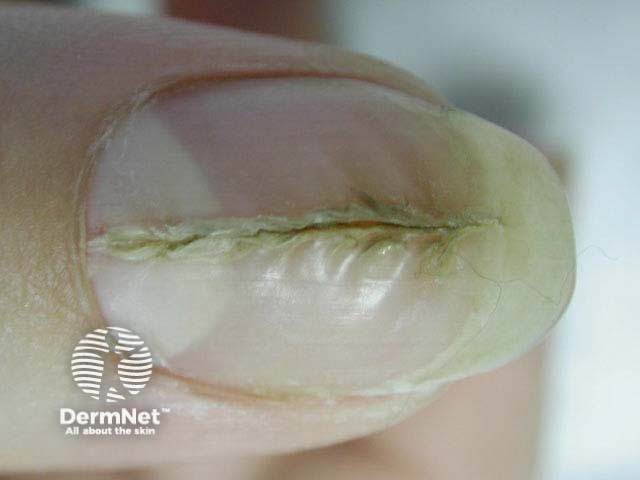
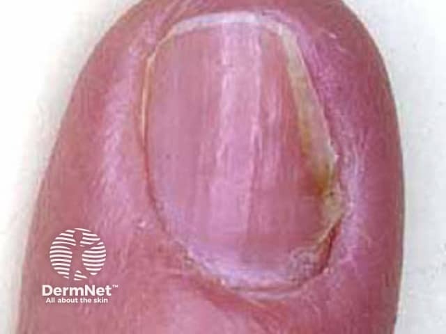
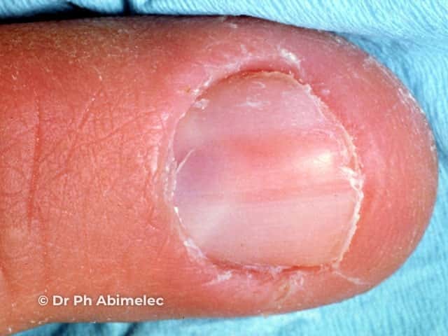
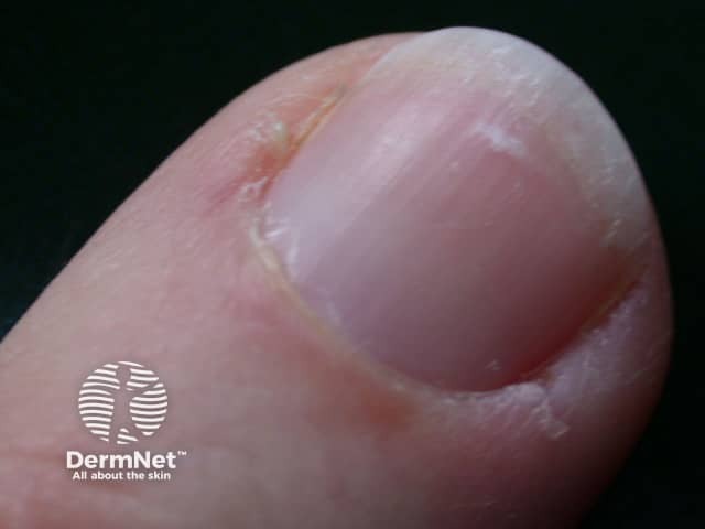
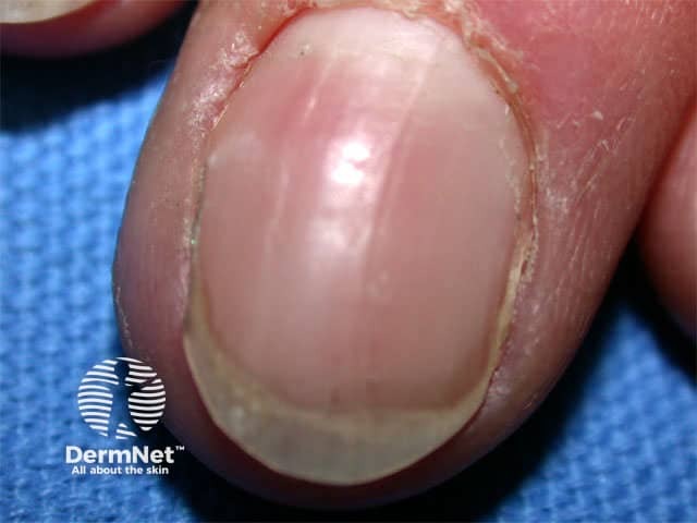
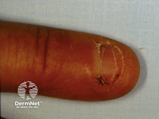
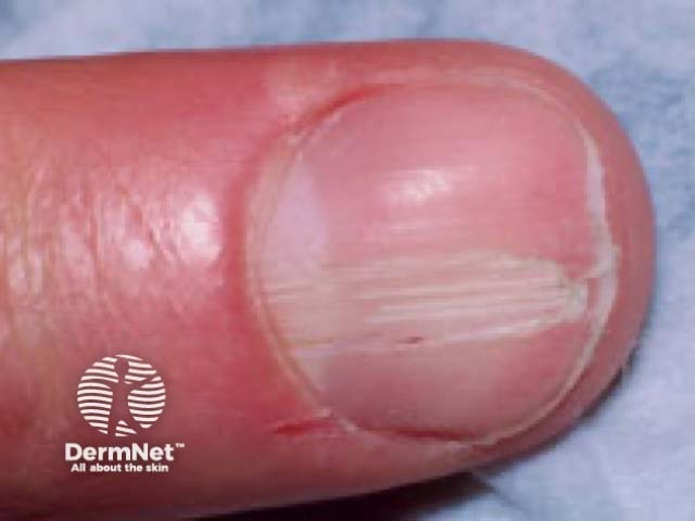
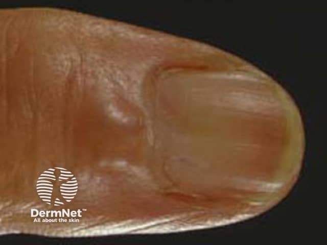
Median canaliform nail dystrophy presents as a feathered, central, longitudinal ridge with a fir-tree pattern usually involving both thumbnails. It is similar to the deformity caused by repetitively pushing back the cuticle (habit-tic deformity). Macrolunulae (large half-moons) may expose the nail matrix to trauma, as they are frequently present.
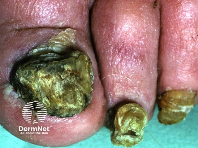
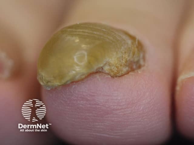
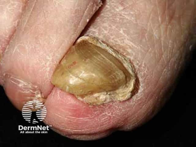
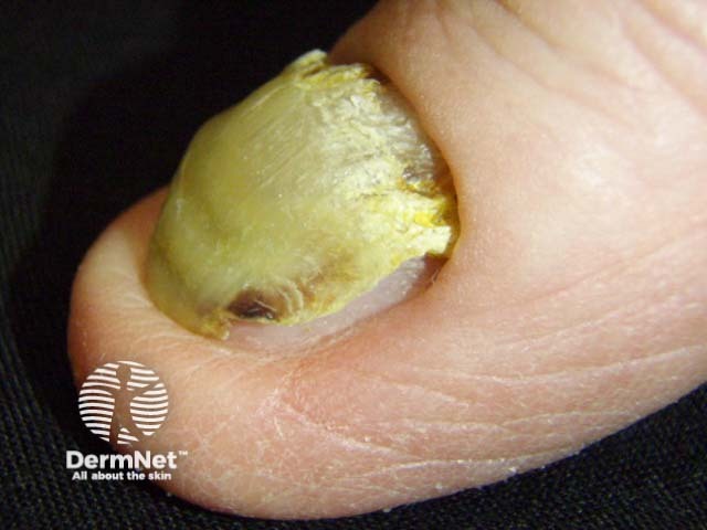
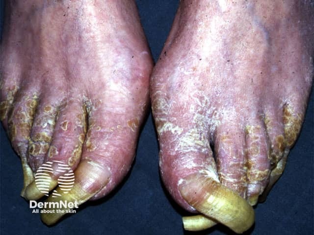
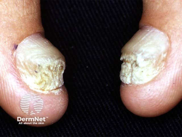
Onychogryphosis is a thick hard curved nail plate in the shape of a ram's horn usually due to ageing, psoriasis or trauma.
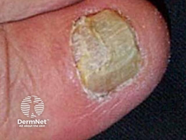
Onychauxis is a thick nail due to psoriasis, trauma or fungal nail infection.
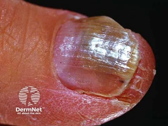
Angel-wing deformity describes nail plate thinning due to lichen planus.
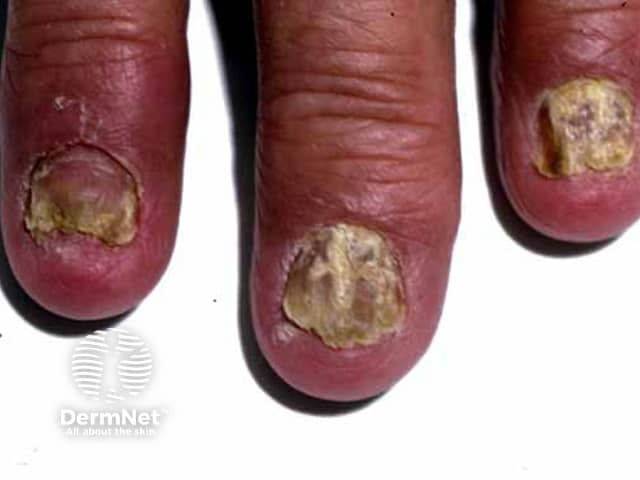
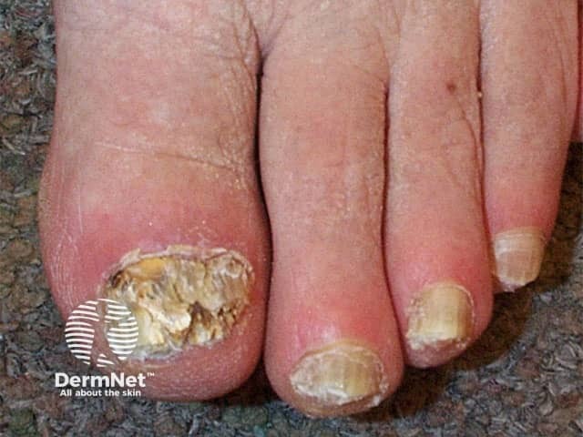
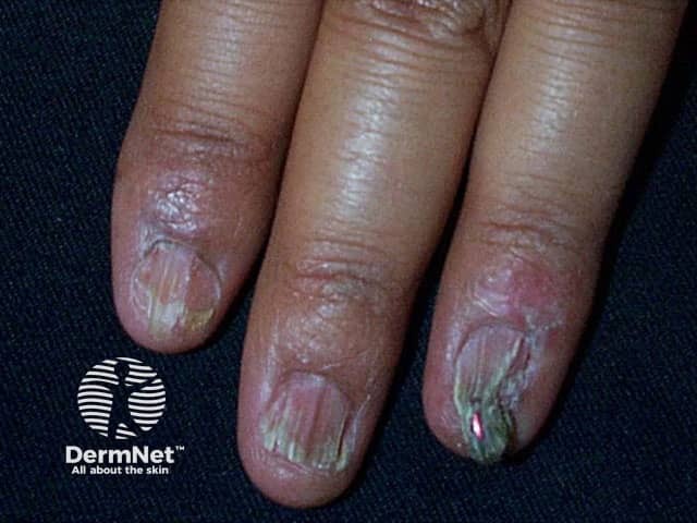
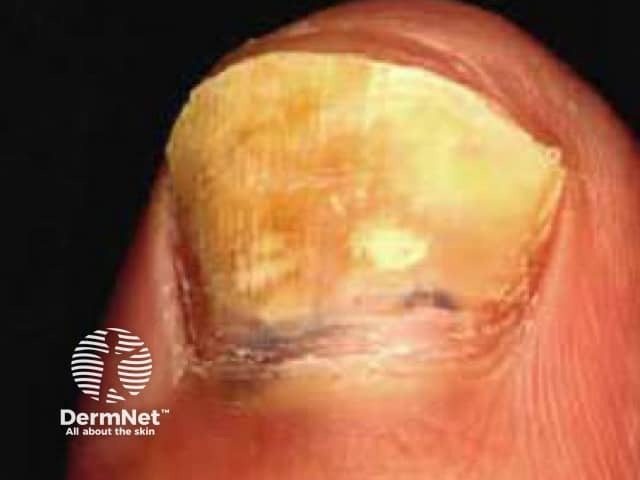
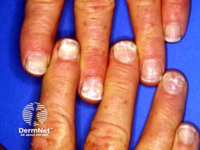
Nail plate crumbling is typical of psoriasis and fungal nail infection.
Onychoschizia is distal lamellar or splitting/brittle nails due to water/detergent damage.
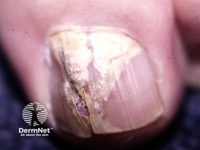
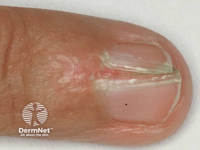
Longitudinal nail splitting is an extension of ridging seen in psoriasis, a fungal nail infection or lichen planus. Distal splitting in association with a pigmented or red linear band can be a sign of onychopapilloma.
Distal notching of the nail occurs from trauma, Darier disease, and lichen planus.
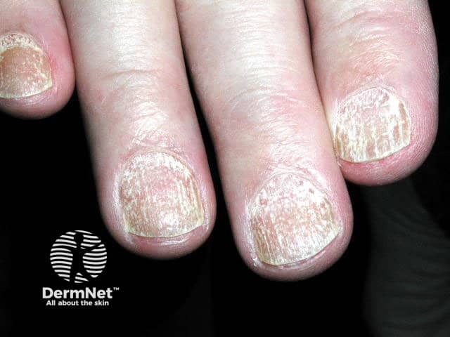
Trachyonychia means rough nails. Trachyonychia is characteristic of lichen planus. Twenty nail dystrophy is trachyonychia of all nails.
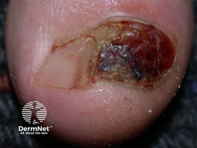
Nail erosion is due to trauma or malignant tumours, such as squamous cell carcinoma or melanoma.
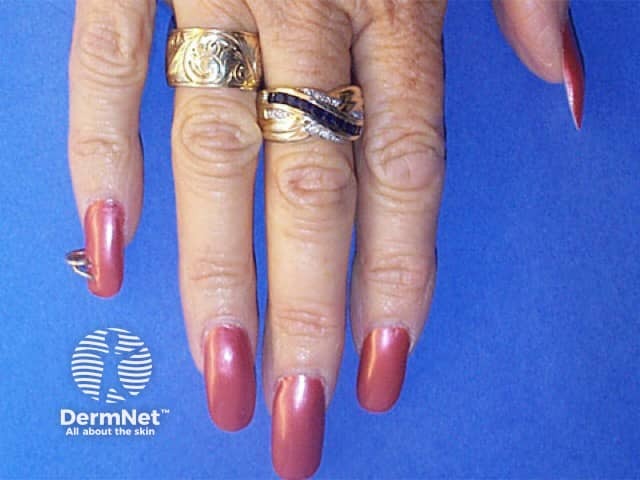
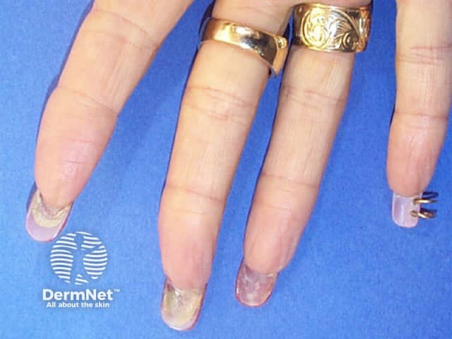
These images show acrylic nails used as decorative cosmetics.
Distinguish a discoloured nail bed from a discoloured nail plate.
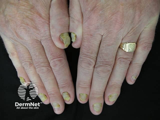
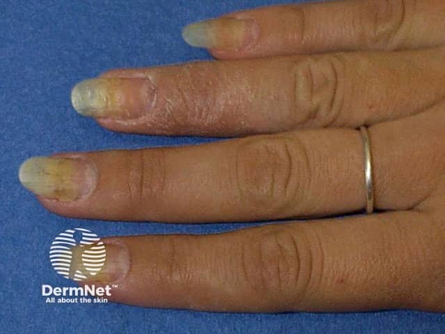
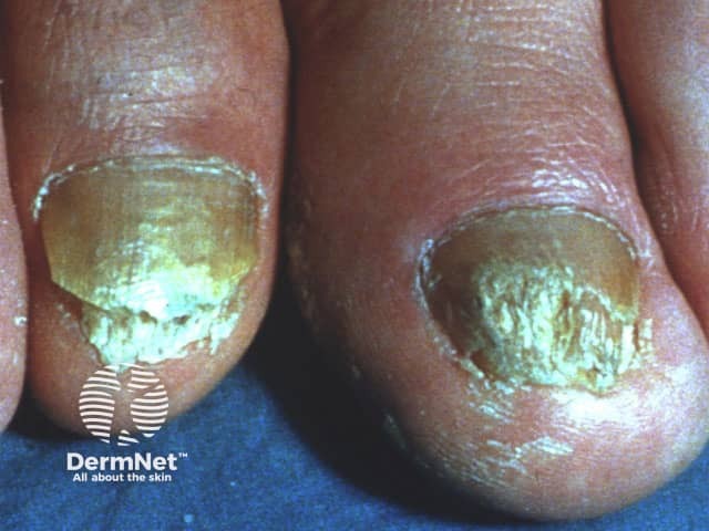
Yellow nail syndrome refers to multiple yellow nails due to lymphatic obstruction in cardiopulmonary disease.
Green nails are associated with Pseudomonas or Candida infection.
See onychomycosis.
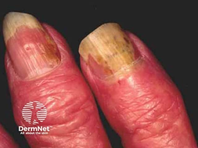
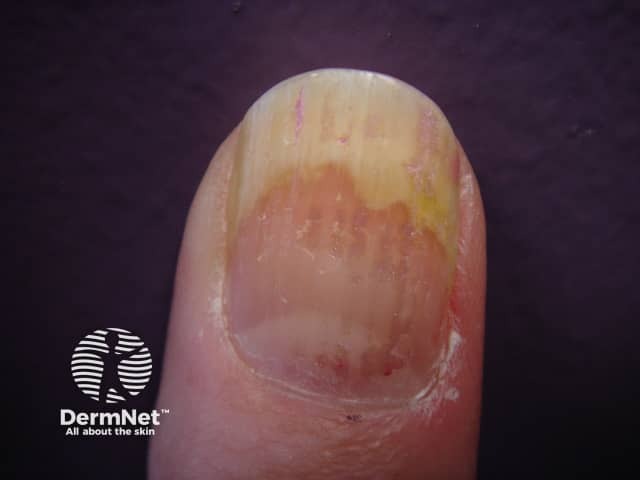
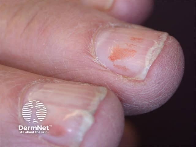
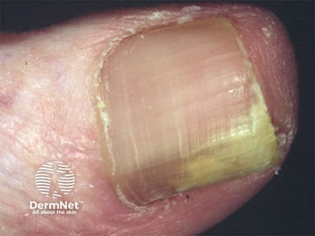
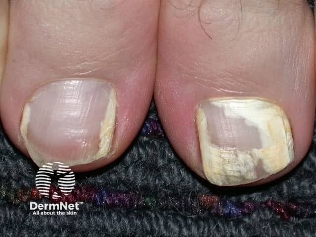
Yellow nails in psoriasis is due to onycholysis, lifting of the nail plate from the nail bed (see below).
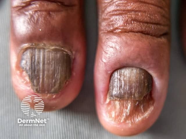
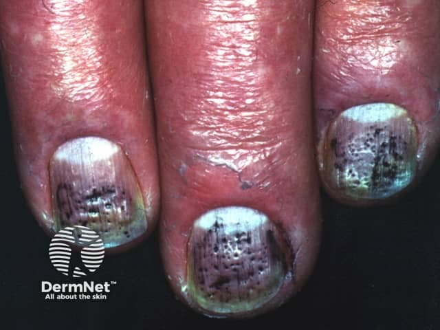
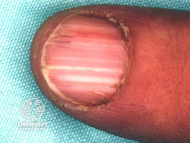
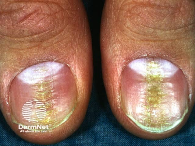
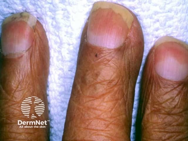
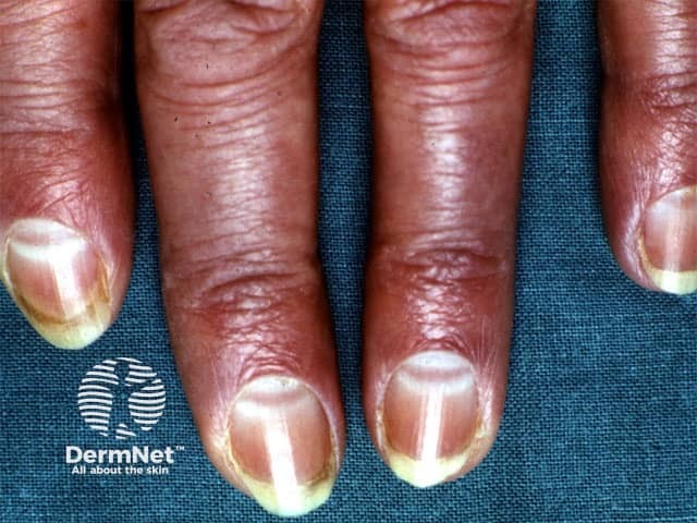
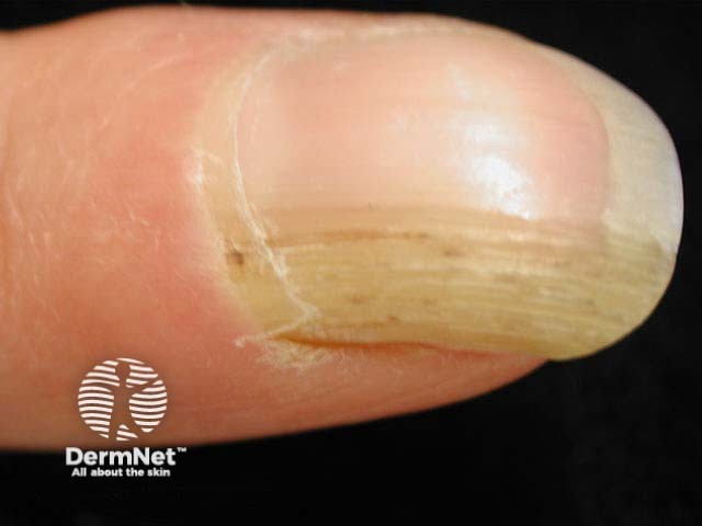
If nails are brown coloured, consider staining (nicotine, potassium permanganate, nail varnish) and chemotherapy. Illustrated are staining from podophyllin and streaks due to oral hydroxyurea. Brown nails may also be due to onychomycosis and in dark-skinned individuals, inflammatory conditions like lichen planus. See also melanonychia and multiple brown linear streaks below.
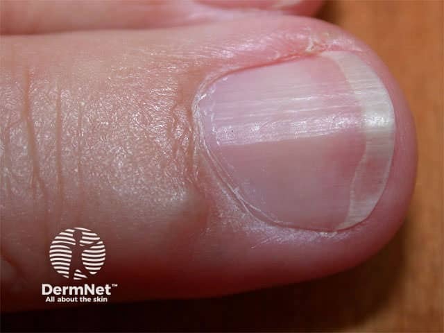
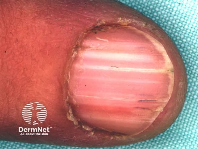
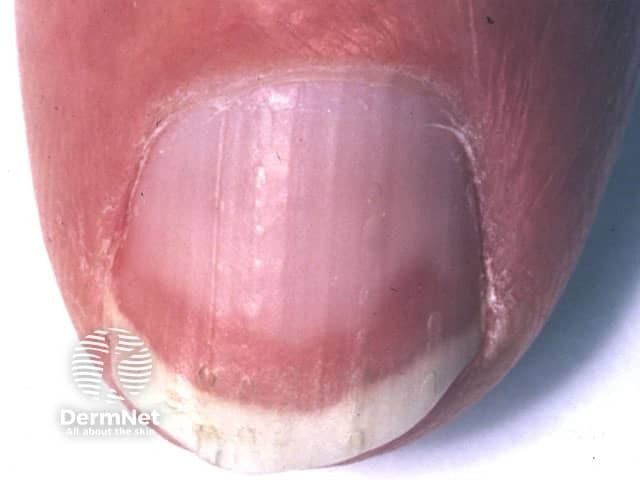
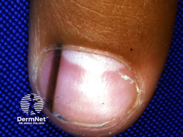
White nails. Consider hypoalbuminaemia or chronic renal failure. May also be familial. Transverse leukonychia, in which there are multiple parallel white lines, is thought to be due to manicuring. It may also arise in association with Beau lines.
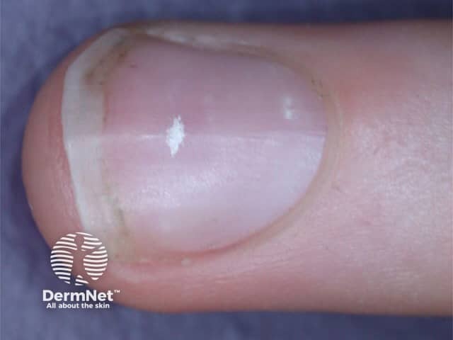
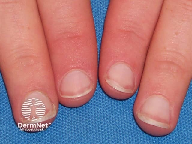
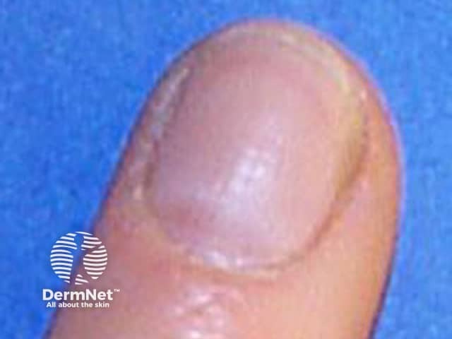
These images show white streaks due to trauma, such as manicuring.
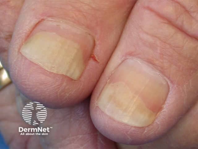
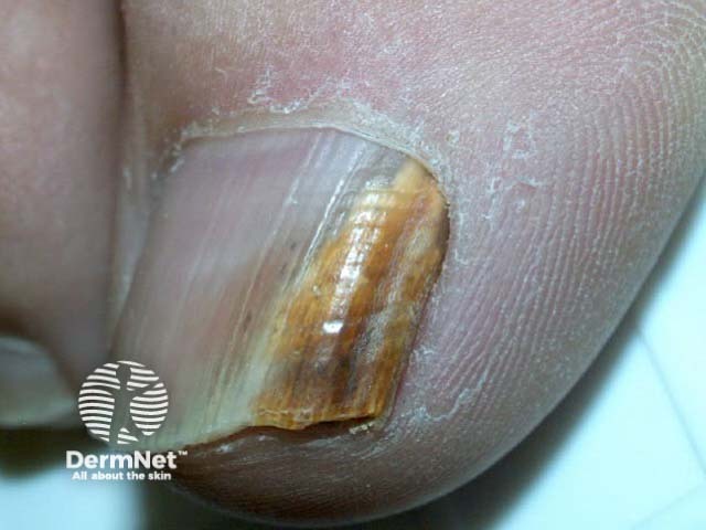
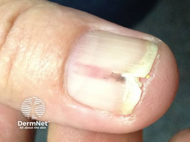
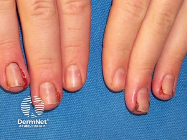
These images show the lifting of the distal nail plate, which appears white or yellow. Consider idiopathic causes, trauma, psoriasis, thyrotoxicosis, irritant and allergic contact dermatitis, fungal nail infection (candida), drug photosensitivity (especially tetracycline and psoralens)
Superficial white onychomycosis
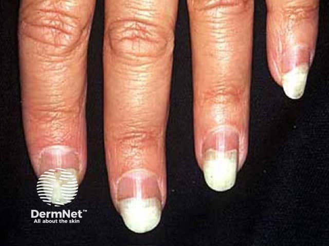
Half-and-half nails occur in renal failure as a white proximal nail, with a brown distal nail.
Terry nail occurring with liver cirrhosis presents as a white proximal nail, and reddened distal nail.
Mee lines are partial leukonychia due to arsenic intoxication or systemic disease.
Muehrcke lines are a double band of leukonychia in renal disease.
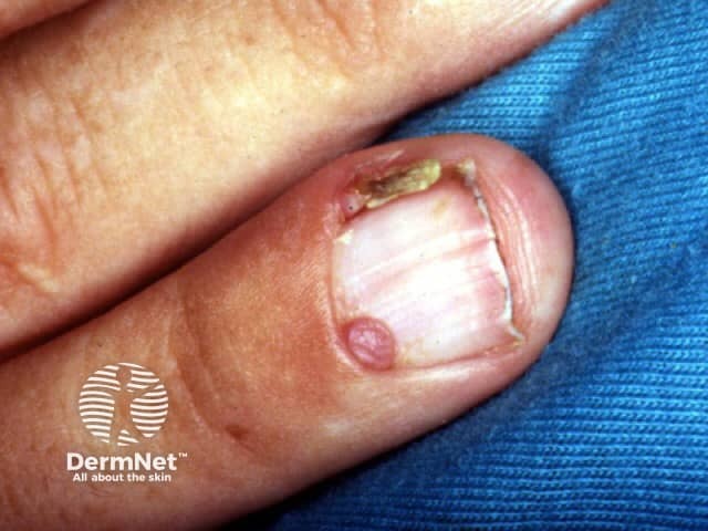
A red longitudinal streak or erythronychia is often due to onychopapilloma.
See Darier disease.
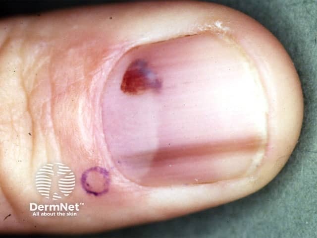
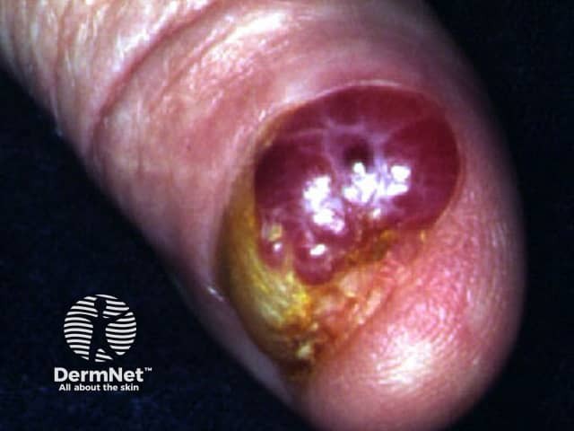
If a red spot is tender, consider glomus tumour.
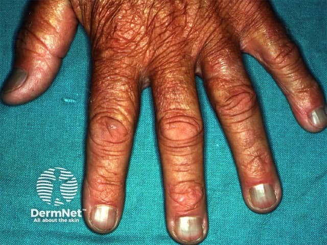
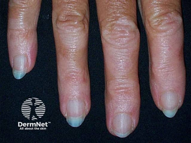
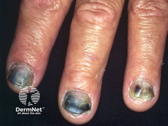
If nails are discoloured blue, consider drugs if all nails are affected, in this case, due to minocycline.
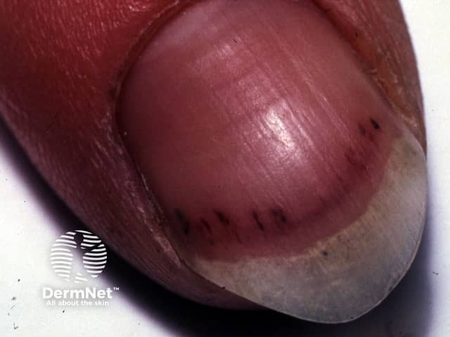
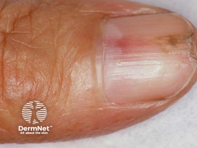
A red or purple streak is known as a splinter haemorrhage.
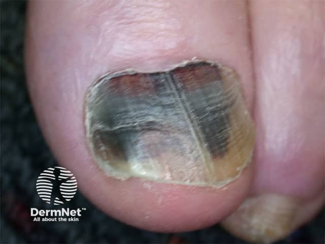
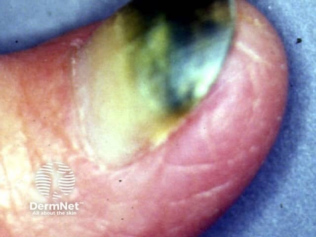
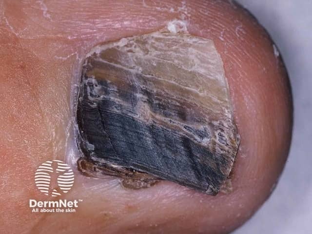
Subungual haemorrhage or haematoma (blood clot) causes a purple or black discolouration.
A black nail may be due to a pseudomonas infection
Melanonychia is a brown/black longitudinal band due to benign melanocytic naevus.

Longitudinal melanonychia may be of racial origin, Laugier-Hunziker syndrome. Rarely due to drug (azidothymidine, tetracycline), endocrine disorders or Peutz-Jeghers syndrome.
Consider melanoma.
The cuticle is a sheet of keratin joining the skin of the proximal nail fold to the nail plate, protecting the space under the proximal nail fold. Loss of cuticle results in paronychia.
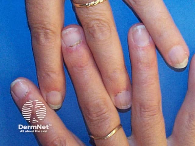
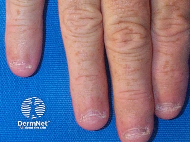
Ragged cuticles are characteristic of connective tissue disease, and also occur in parakeratosis pustulosa.
Hangnail can be due to trauma (eg, nail biting).
Nail fold telangiectases are characteristic of connective tissue disease, for example, lupus erythematosus.
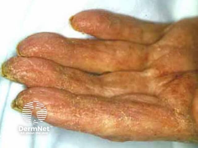
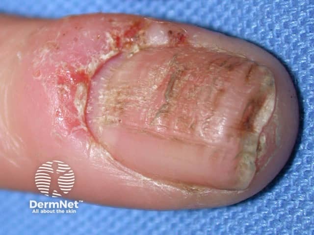
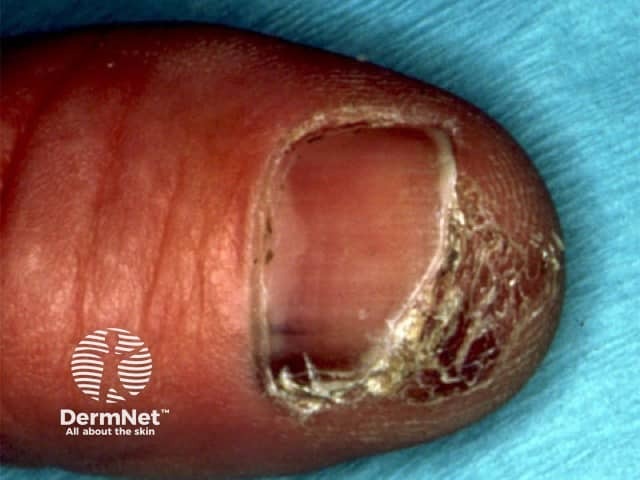
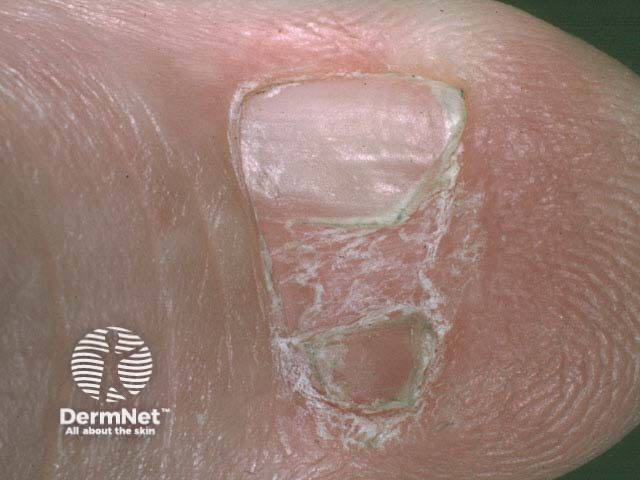
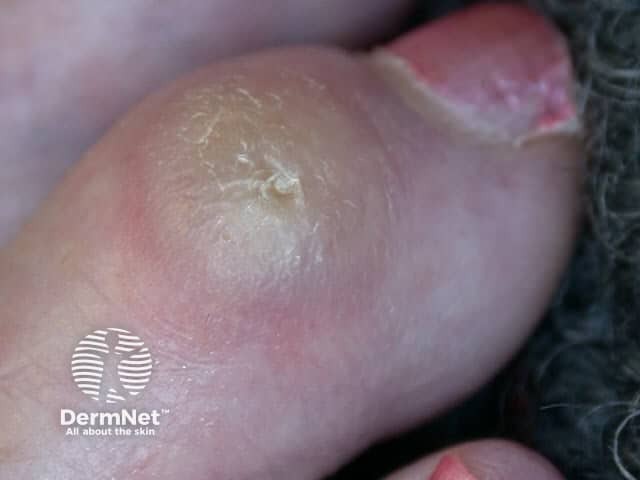
Subungual hyperkeratosis is scaling under the hyponychium. Typical of psoriasis and onychomycosis, it is also seen in crusted scabies.
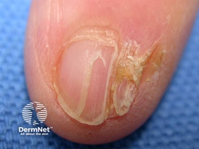
A pterygium is a wing of extra tissue. In a nail, it is due to scarring in the matrix. Characteristic of lichen planus, but may also occur in Stevens-Johnson syndrome and after trauma.
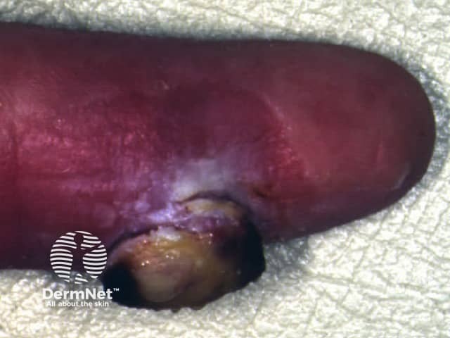
Staphylococcus aureus is the main cause of acute paronychia.
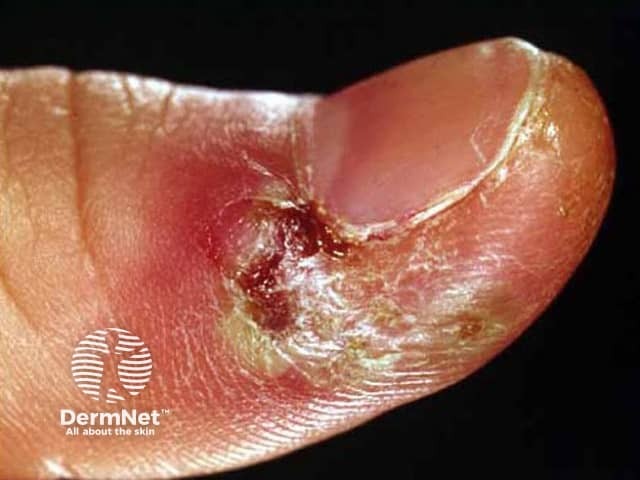
Herpes simplex is another common cause of acute paronychia (see Herpetic whitlow).
Chronic paronychia is associated with dermatitis, and infection with Candida albicans and Pseudomonas.
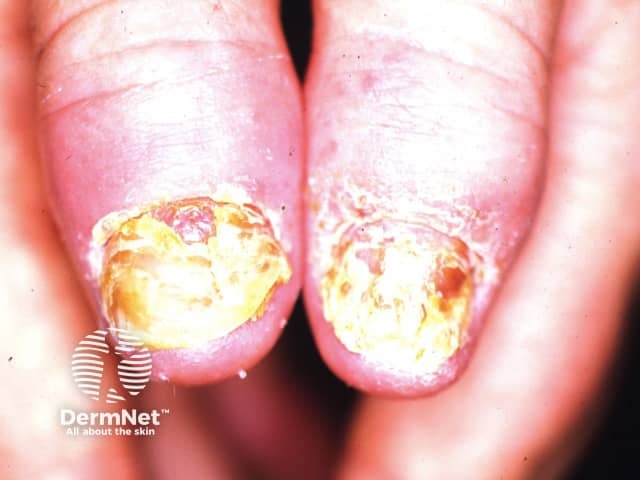
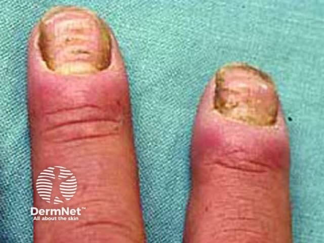
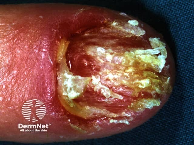
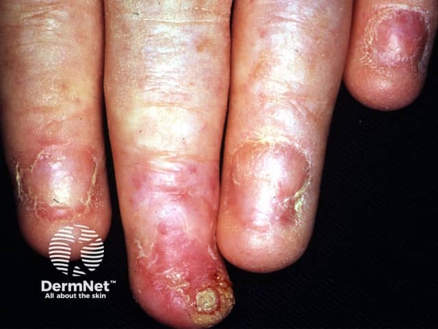
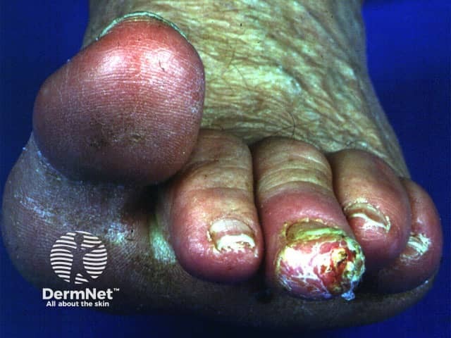
Acrodermatitis continua of Hallopeau is a form of severe psoriasis
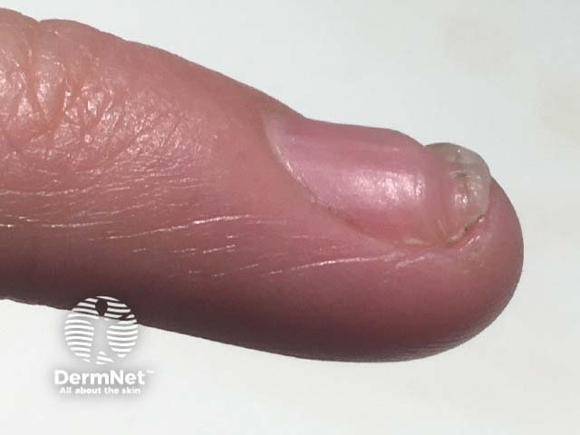
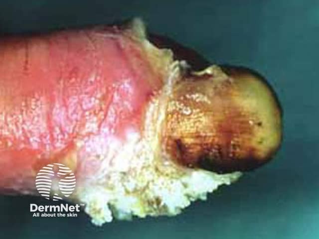
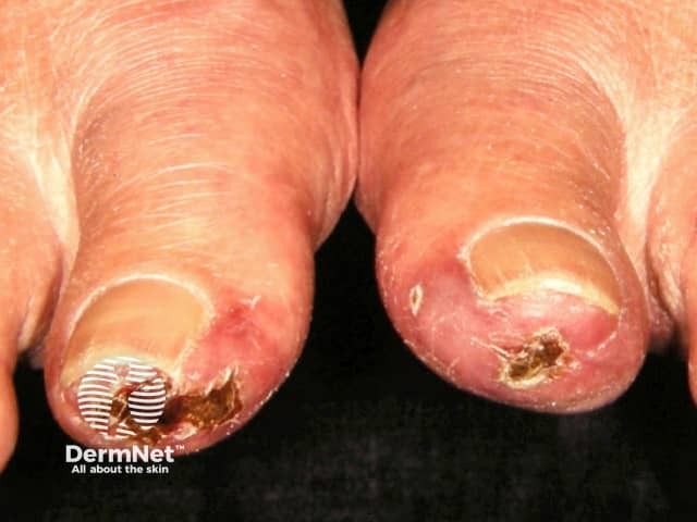
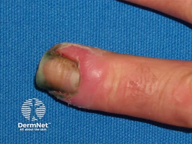
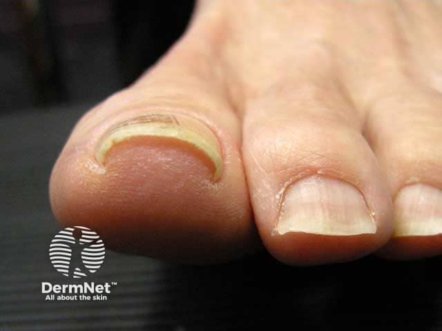
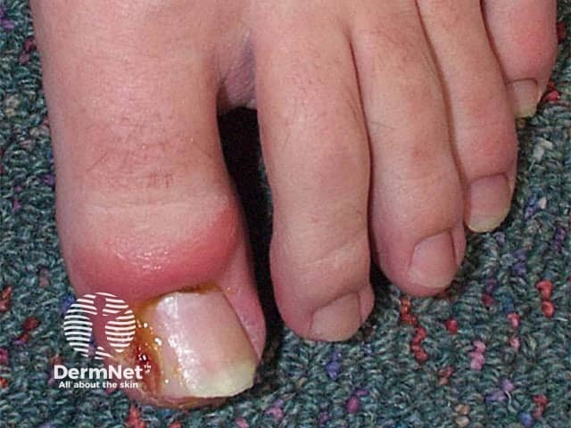
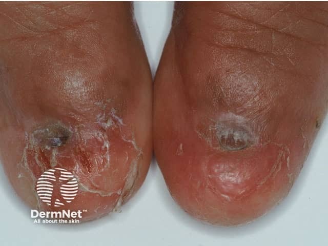
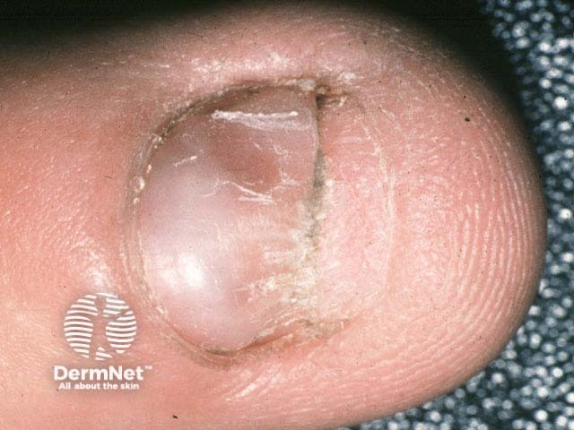
Retronychia refers to the embedding of the proximal nail plate into the proximal nail fold with subsequent painful nail fold inflammation and thickening, and granulation tissue, usually seen in the great toes. It typically results from trauma pushing the nail plate up with a new plate growing out underneath. Treatment is the removal of the nail.
Short nail
Clubbing is due to hypertrophic osteoarthropathy or thyroid disease (acropachy); see hypertrophic osteoarthropathy.
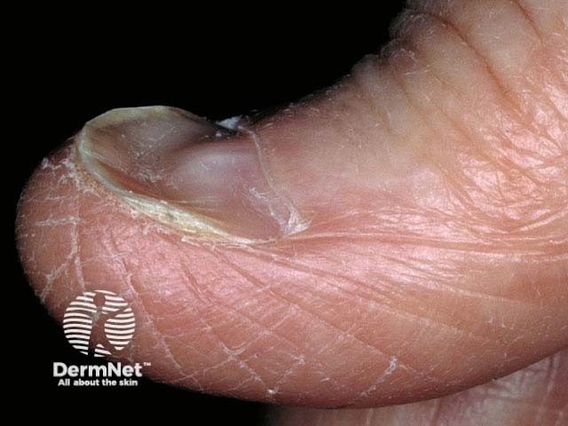
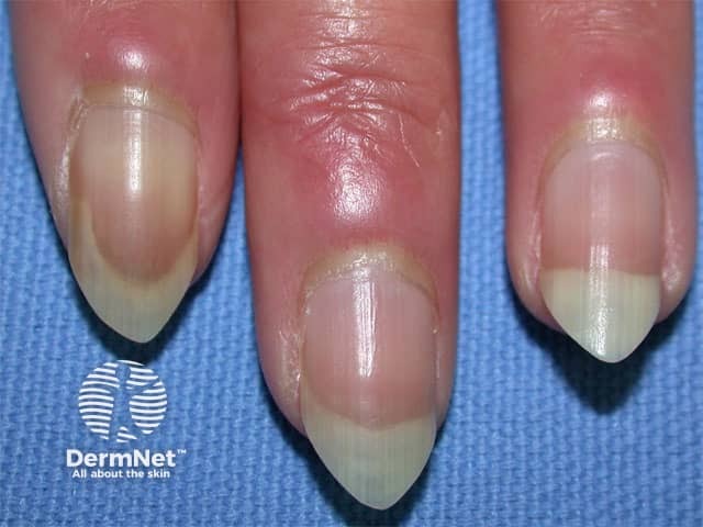
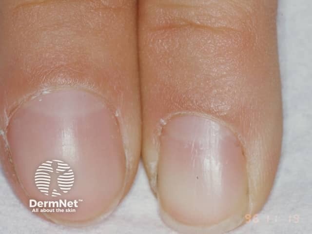
Koilonychia is a thin, spoon-shaped nail, and can occur in normal children and adults. It is also associated with iron deficiency anaemia, diabetes, protein deficiency, connective tissue disease, nail exposure to solvents and acitretin treatment.
Pincer nail is sometimes familial or associated with psoriasis.
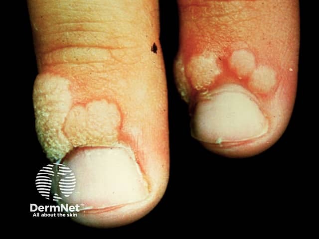
Onychocryptosis is commonly known as an in-grown nail, and involves the lateral nail folds with granuloma formation. Often aggravated by oral retinoids, isotretinoin and acitretin.
Usually, traumatic, such as nail-biting; in children parakeratosis pustulosa
Common skin lesions around nails include:

An AI summary will appear based on your search term using data from all of the topic pages across the entire DermNet site.
Show more