Author: Dr Diana Purvis, Dermatologist, Starship Hospital, Auckland, New Zealand; Chief Editor: Hon A/Prof Amanda Oakley, Dermatologist, Hamilton, New Zealand, December 2016. Minor update by Ian Coulson, Dermatologist. Copy edited by Gus Mitchell. July 2024.
Onychopapilloma is a benignneoplasm of the nail matrix. It is usually an isolated lesion affecting one nail. Multiple nail involvement is rare and may be a sign of a BAP1 tumourpredispositionsyndrome. Rarely malignant onychopapilloma may occur with a histology of Bowen’s disease.
Who gets onychopapilloma?
Onychopapiloma is rare. It can affect males and females of all races and ethnicities. It has mainly been described in middle-aged and older adults.
What causes onychopapilloma?
The cause of onychopapilloma is unknown. Multiple polydactylous lesions have been associated with the BAP1 tumour predisposition syndrome. In this situation individuals have a higher risk of multiple Spitz tumours, ocular and cutaneousmelanoma, clear cell carcinoma of the kidney and mesothelioma.
What are the clinical features of onychopapilloma?
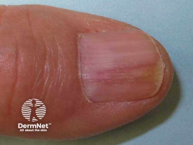
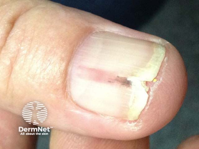
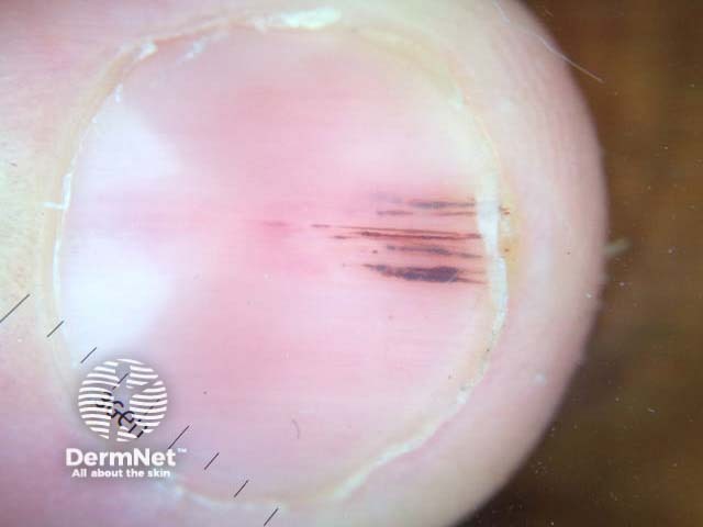
Onychopapilloma usually results in a longitudinal streak in a red nail (erythronychia), which extends from the lunula to the tip of the nail.
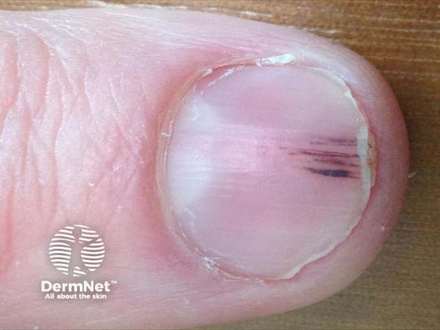
White longitudinal streaks (leukonychia) or brown longitudinal streaks (melanonychia) may also occur.
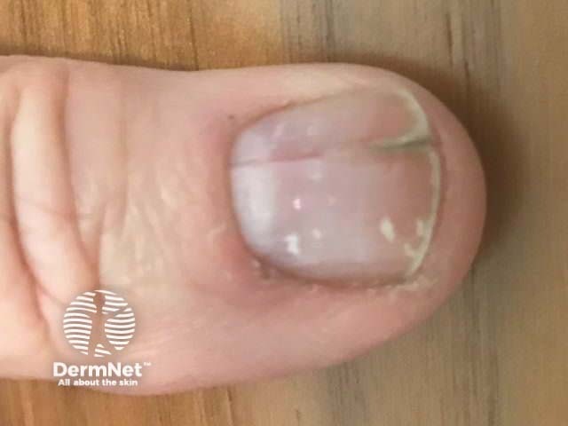
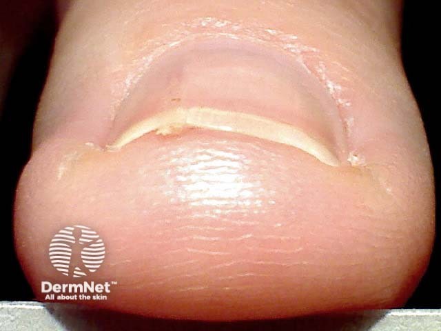
The distal tip of the nail may split or lift (onycholysis).
A warty growth may be evident under the nail (subungualkeratosis).
Dermoscopy of the free edge of the nail plate shows a small area of subungual scale.
Erythronychia, onycholysis
Splinter haemorrhages
Melanonychia
Erythronychia
Dermoscopy
Subungual hyperkeratosis
How is onychopapilloma diagnosed?
The diagnosis may be suspected clinically, but as malignant lesions under the nail could look similar, it is important to keep the lesion under review and undertake nail biopsy if there is an enlarging lesion.
Histology of a nail clipping showing an area of asymmetricaldyskeratosis and papillomatosis.
Longitudinal biopsy of the nail shows a papillomatous nail bed with layers of hyperkeratosis and an absent granular layer.Pigmentation if present, is due to melanocyte activation.
What is the differential diagnosis for onychopapilloma?
The differential diagnosis for onychopapilloma includes:
Malignant onychopapilloma - widening of the longitudinal band and pain may alert to this entity.
What is the treatment for onychopapilloma?
Longitudinal excision of the entire affected nail and proximalnail matrix is curative.
What is the outcome for onychopapilloma?
Untreated, the lesion tends to persist.
References
Tosti A, Schneider SL, Ramirez-Quizon MN, Zaiac M, Miteva M. Clinical, dermoscopic, and pathologic features of onychopapilloma: A review of 47 cases. J Am Acad Dermatol. 2016 Mar;74(3):521–6. doi: 10.1016/j.jaad.2015.08.053. Review. PubMed PMID: 26518173. PubMed.
Min, JungChae, Seoung WanLee, Ga-Young et al. Case of onychopapilloma presenting as longitudinal melanonychia. Dermatologica Sinica, Volume 0, Issue 0. Full text.
Baran R, Perrin C. Longitudinal erythronychia with distal subungual keratosis: onychopapilloma of the nail bed and Bowen's disease. Br J Dermatol. 2000 Jul;143(1):132–5. PubMed PMID: 10886147. PubMed.
Lebensohn A, Ghafoor A, Bloomquist L, et al. Multiple Onychopapillomas and BAP1 Tumor Predisposition Syndrome. JAMA Dermatol. Published online May 17, 2024. doi:10.1001/jamadermatol.2024.1804. PubMed
Haneke E, Iorizzo M, Gabutti M, Beltraminelli H. Malignant onychopapilloma. J Cutan Pathol. 2021;48(1):174–9. PubMed