Herpetic whitlow is a painful viral cutaneousinfection that usually affects the distal fingers or thumbs, and occasionally the toes. It is caused by herpes simplex virus (HSV) type 1 or 2, and can be vesicular or pustular in nature.
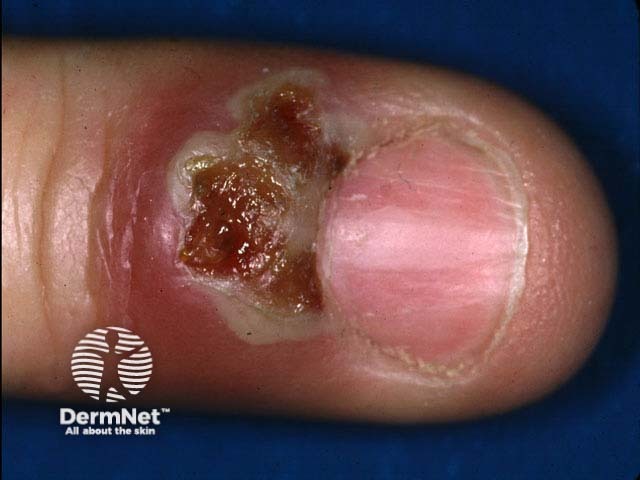
Clustered vesicles, due to HSV, have broken down into erosions on the proximalnail fold
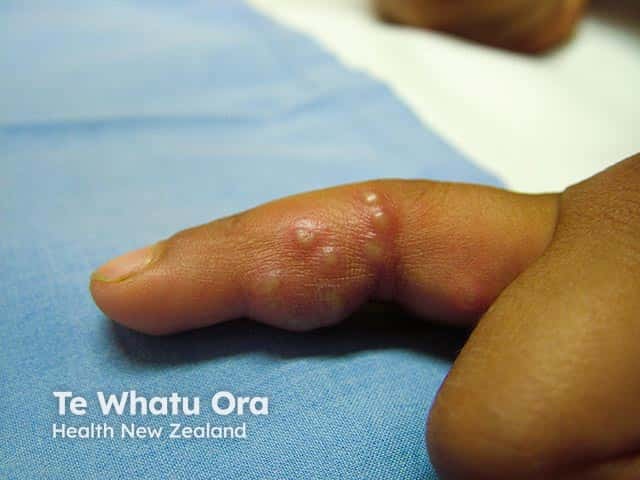
Several nail folds are affected by herpetic whitlow in a woman receiving chemotherapy
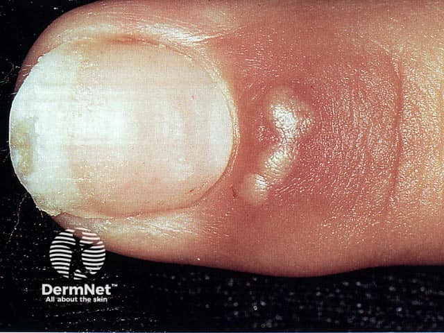
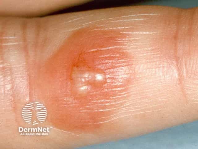
Clustered clear vesicles on the proximal nail fold typical of an early phase of a herpetic whitlow
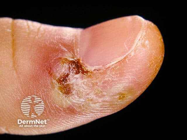
A herpetic whitlow on the thumb five days after onset
Clustered clear vesicles and oedema in an early herpetic whitlow in skin of colour (HW-patient1)
One case series reported the incidence of herpetic whitlow as 2.4 cases per 100,000 people per year. Males and females appear to be equally affected.
In young children, herpetic whitlow commonly occurs following oral herpes infection. This is thought to be related to thumb sucking causing autoinoculation from active herpetic lesions in the mouth.
Herpetic whitlow in adults is more commonly associated with exposure to genital herpes, or occupational exposure of dental and other health care workers to patients with herpes simplex including oral secretions.
People who are immunosuppressed are also more susceptible to herpetic infections.
What causes herpetic whitlow?
Autoinoculation through spread from other herpetic lesions such as oral or genital herpes.
Direct exogenousinoculation from an external source.
Viral reactivation following previous herpes simplex virus (HSV) type 1 or 2. HSV results in viral invasion and replication in epidermal and dermal cells, which can progress to involve the sensory dorsal root ganglion. Here, HSV remains and can be periodically reactivated to cause disease anywhere (including the fingertips).
Patients may report preceding trauma such as a torn cuticle.
What are the clinical features of herpetic whitlow?
Patients typically present with a painful swollen digit, often the thumb or index finger.
Possible prodromalfever and malaise.
Localised itch, burning, and altered sensation (may precede vesicle formation).
One or more (often clustered) 1–3 mm fluid-filled vesicles with surrounding erythema, most often affecting the distal phalanx.
Vesicular fluid is usually clear but can progress to become turbid, sero-purulent, or haemorrhagic.
Vesicles can coalesce into larger bullae and can spread proximally.
Proximal lymphangitis and lymphadenopathy may be observed.
Typically, the vesicles will crust and desquamate over several weeks and resolve without any treatment.
Other herpetic lesions (eg, oral or genital) may also be present.
The thumb or index finger are most commonly affected; it is unusual for more than one digit to be affected.
Superimposed bacterial or fungal infection and potential abscessdevelopment.
Secondary herpes simplex infection including spread to oral, ocular, or genital areas.
Recurrence (~23% in one paediatric review).
How is herpetic whitlow diagnosed?
Herpetic whitlow can be diagnosed clinically based on history and appearance of the lesions.
Viral cultures or a Tzanck smear can be used to confirm the diagnosis. Ballooning multinucleatedgiant cells and eosinophilicinclusion bodies are seen on the Tzanck smear. A bacterial swab can also be taken if secondary or concurrent bacterial infection is suspected.
What is the differential diagnosis for herpetic whitlow?
Early localised hand, foot, and mouth disease (HFMD) can present similarly with vesicular or papulovesiculareruption.
What is the treatment for herpetic whitlow?
General measures
Education on skin protection — keep affected digit/s clean and covered with a dressing to prevent further irritation and spread, as viral shedding can occur until all lesions have cleared.
Antivirals (eg, topical or oral aciclovir, or valaciclovir) commenced within 48 hours of symptom onset may reduce the duration of symptoms and risk of recurrence.
Consider suppressive or prophylacticantiviral therapy for those with recurrent flares or severe episodes, especially if they are immunosuppressed.
For superimposed bacterial infection, antibiotics may be indicated.
Surgical drainage is not recommended due to the risk of viraemia and secondary bacterial infection.
Consider testing for HIV if the lesion is extensive and if there are risk factors.
How can herpetic whitlow be prevented?
Those at higher occupational risk are advised to use gloves when in contact with patients with symptoms of herpes simplex.
Children with oral herpes should be discouraged from sucking their thumb.
What is the outcome for herpetic whitlow?
Herpetic whitlow is self-limiting and usually resolves without any complications in 2–4 weeks. Recurrence is possible due to reactivation of the virus, which can be triggered by stress, other illness, or trauma to the skin or nails. Recurrent episodes are usually less severe than the primary infection.
Betz D, Fane K. Herpetic Whitlow. In: StatPearls. Treasure Island (FL): StatPearls Publishing; 2022. Available here
Gill MJ, Arlette J, Buchan K. Herpes simplex virus infection of the hand: A profile of 79 cases. Am J Med. 1988;84(1):89–93. doi: 10.1016/0002-9343(88)90013-7. Journal
Klotz RW. Herpetic whitlow: an occupational hazard. AANA J. 1990;58(1):8–13. Journal
Lieberman L, Castro D, Bhatt A, Guyer F. Case report: palmar herpetic whitlow and forearm lymphangitis in a 10-year-old female. BMC Pediatr. 2019;19(1):450. doi: 10.1186/s12887-019-1828-5. Journal
Robayna MG, Herranz P, Rubio FA, et al. Destructive herpetic whitlow in AIDS: report of three cases. Br J Dermatol. 1997 Nov;137(5):812–5. Journal
Szinnai G, Schaad UB, Heininger U. Multiple herpetic whitlow lesions in a 4-year-old girl: case report and review of the literature. Eur J Pediatr. 2001 Sep;160(9):528–33. doi: 10.1007/s004310100800. Journal