
ADVERTISEMENT
You do not have any notes added to this page yet
Introduction Demographics Causes Clinical features Complications Diagnosis Differential diagnoses Treatment Outcome
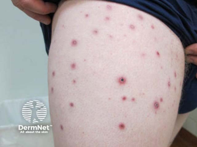
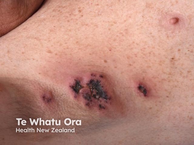
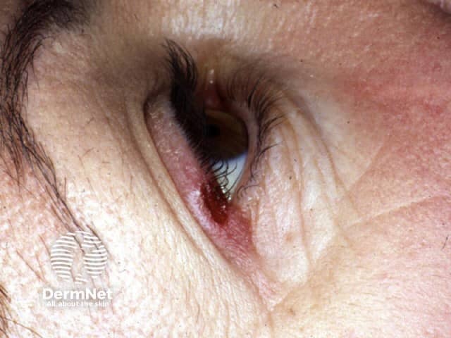
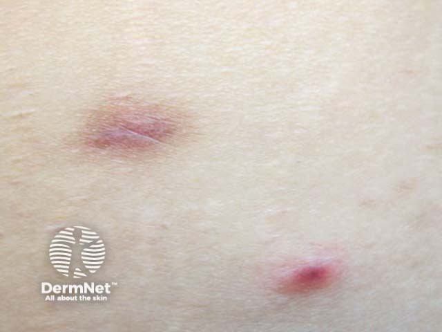
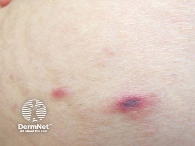
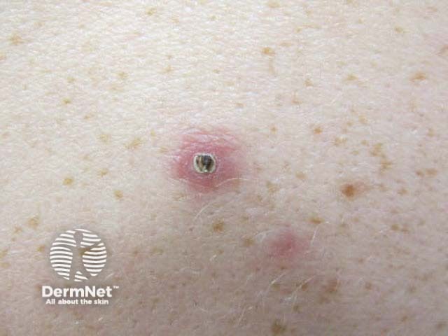
Lymphomatoid papulosis (LyP) is a rare form of indolent cutaneous T-cell lymphoma characterised by crops of recurrent self-healing papules. It is the most common type of primary cutaneous CD30+ lymphoproliferative disorder.
Lymphomatoid papulosis has an estimated incidence of 1.2–1.9 cases per million population. LyP can affect all races, although it is uncommon in skin of colour. Any age can be affected; the average age of onset is between 35 and 45 years. However, even children can present with lymphomatoid papulosis. Men are more commonly affected than women.
The aetiopathogenesis of primary cutaneous CD30+ lymphoproliferative disorders remains elusive. CD30 signalling is known to affect growth and survival of lymphoid cells. Some studies suggest restriction of the normal T cell repertoire in the peripheral blood. Other hypotheses implicate reactive phenomena inducing CD30+ overexpression.
Lymphomatoid papulosis can be associated with other cutaneous or systemic lymphomas which may precede, accompany, or follow the diagnosis of LyP.
Quality of life can be impacted by cosmetically-troubling LyP lesions and scarring.
Diagnosis of lymphomatoid papulosis requires careful clinicopathological correlation. History, examination, and skin histopathology must be compatible. The diagnosis cannot be made on histology alone as similar findings can be seen in other skin conditions.
Imaging eg, CT or PET scans, and bone marrow examination is not indicated unless there are clinical features to suggest extracutaneous disease.
Currently there is no curative treatment for lymphomatoid papulosis, nor treatments that change the natural course of the disease, although healing can be accelerated and the frequency and/or severity of crops reduced by some therapies. A wait-and-see approach can be considered given the indolent course and common relapse after ceasing active therapies.
The choice of treatment and escalation of therapy should be made by weighing any potential risks against short-term benefits. Selecting an appropriate treatment modality will depend on a number of factors including, but not limited to, the extent of lesions, frequency of flares, and involvement of cosmetically sensitive or highly visible areas.
Despite the malignant appearance on histology, lymphomatoid papulosis typically has an indolent waxing and waning course lasting months or even decades before remitting. The five-year survival is estimated to be 100%.
Long-term follow-up and surveillance is required as up to 20% of cases progress to other forms of cutaneous or systemic lymphoma such as mycosis fungoides, Hodgkin lymphoma, primary cutaneous anaplastic large-cell lymphoma, or nodal anaplastic large-cell lymphoma. Risk factors for associated lymphoma include older age and the presence of a monoclonal TCR-gene rearrangement on skin biopsy. Progression to a second or third lymphoma can occur after a prolonged delay of five or more years.
An AI summary will appear based on your search term using data from all of the topic pages across the entire DermNet site.
Show more