Authors: Vanessa Ngan, Staff Writer, 2003. Updated: Dr Maanasa Bandla, Intern, Monash Health, Melbourne, Australia; Dr Martin Keefe, Dermatologist, Christchurch, New Zealand. Copy edited by Gus Mitchell. August 2021
Folliculitis decalvans is a chronic, neutrophilicinflammation that results in scarring hair loss. Tufted hair folliculitis is probably a subset of folliculitis decalvans although tufting can be seen in other forms of cicatricialalopecia as well.
Folliculitis decalvans
Folliculitis decalvans
Who gets folliculitis decalvans?
Folliculitis decalvans usually presents in the 4th and 5th decades, with a male predominance. Children are not affected.
There are probably no racial differences in prevalence, although it has been claimed to be more common in African-American women.
Familial cases have been rarely reported.
What causes folliculitis decalvans?
Folliculitis decalvans is considered to be the result of an abnormal immune response to Staphylococcus aureus, although this is not yet proven.
What are the clinical features of folliculitis decalvans?
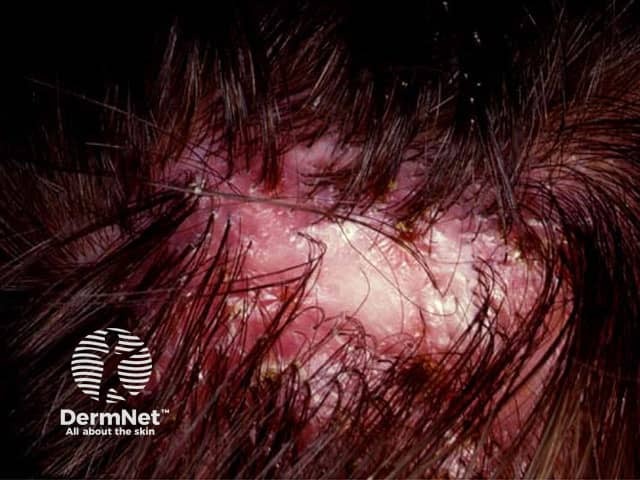
Folliculitis decalvans typically affects the scalp, often around the crown, but may affect the beard area, axillae, limbs, and pubic hair. The characteristic clinical features include:
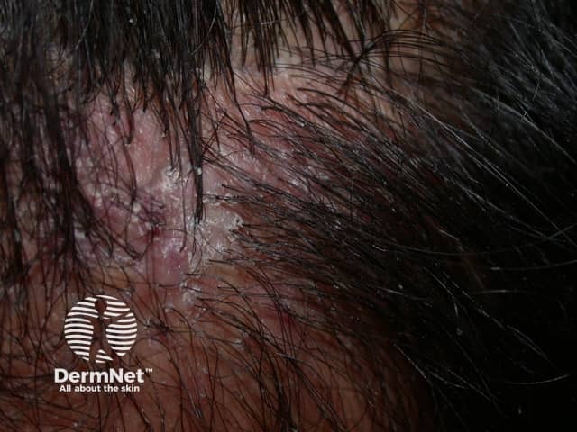
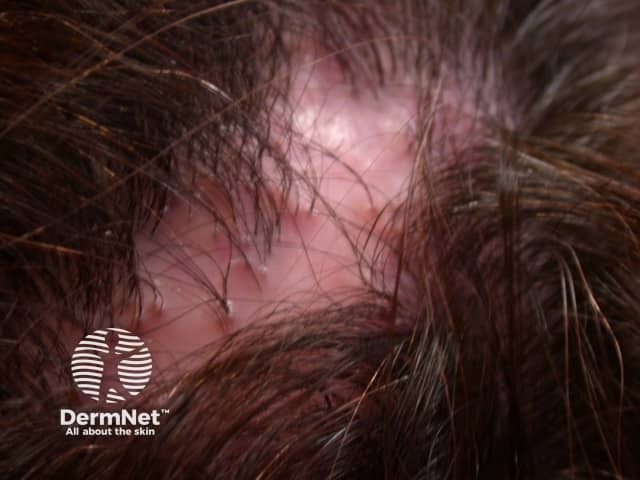
Irregular, atrophic white patches of scarring and hair loss — solitary or multiple
Induration of the scalp
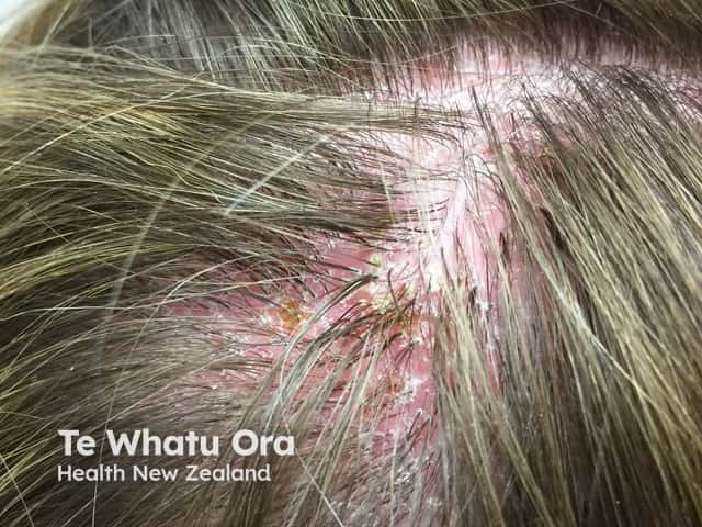
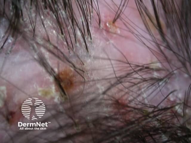
Follicularpustules and perifollicular crusts at the patch periphery
Follicular hyperkeratosis, scale, and erosions
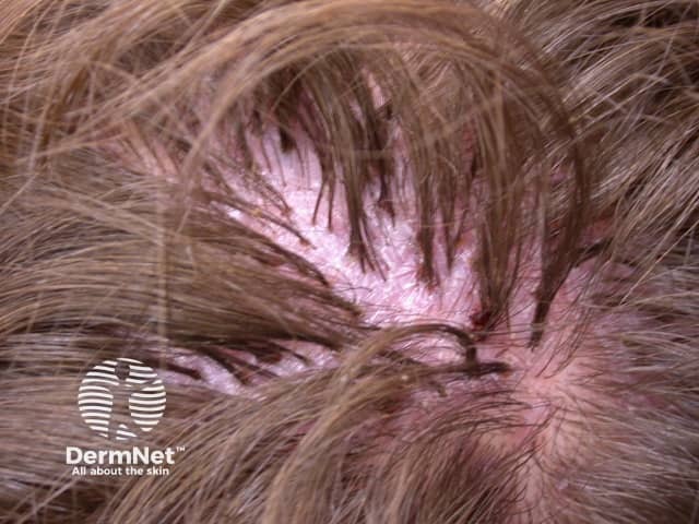
Tufting — multiple hair shafts emerge from a single hair follicle, resulting in a ‘doll’s hair’ appearance
Mild itch, discomfort, or pain.
Perifollicular crusting and scale
Tufted hairs and perifollicular crusting
Tufted hairs and perifollicular erythema
Dermoscopy of folliculitis decalvans
Tufted hairs
White dots
Perifollicular erythema and scale
Scattered follicular pustules
What are the complications of folliculitis decalvans?
Folliculitis decalvans is a clinical diagnosis confirmed on bacteriology/mycology and histology if required.
Skin biopsy of an early lesion shows a neutrophilic infiltrate dilating the infundibulum of the hair follicle. The follicle in later lesions has ruptured resulting in perifollicular scarring and mixed inflammatory infiltrate including foreign body giant cells.
What is the differential diagnosis for folliculitis decalvans?
Folliculitis decalvans usually follows a chronic fluctuating course of exacerbations and remissions over many years. It is not clear that treatment influences the long-term prognosis despite successfully reducing inflammation in the short-term. Early diagnosis and treatment are important but permanent hair loss is to be expected.
Doche, I., Sotto, M. N., Hordinsky, et al. (2025). Hydroxychloroquine may be beneficial as an adjuvant therapy for folliculitis decalvans: a 5-year retrospective study with 49 patients. Clinical and Experimental Dermatology, 50(2), 404–407. doi:10.1093/ced/llae394. Journal
Fässler M, Radonjic-Hoesli S, Feldmeyer L, et al. Successful treatment of refractory folliculitis decalvans with apremilast. JAAD Case Rep. 2020;6(10):1079–81. doi:10.1016/j.jdcr.2020.08.019. PubMed Central
Otberg N, Kang H, Alzolibani AA, Shapiro J. Folliculitis decalvans. Dermatol Ther. 2008;21(4):238–44. doi:10.1111/j.1529-8019.2008.00204.x. PubMed
Rambhia PH, Conic RRZ, Murad A, Atanaskova-Mesinkovska N, Piliang M, Bergfeld W. Updates in therapeutics for folliculitis decalvans: a systematic review with evidence-based analysis. J Am Acad Dermatol. 2019;80(3):794–801.e1. doi:10.1016/j.jaad.2018.07.050. Journal
Senatore S, Maglie R, Maio V, Montefusco F, Antiga E. Folliculitis decalvans with exclusive beard involvement. Indian J Dermatol Venereol Leprol. 2021;87(4):569–71. doi:10.25259/IJDVL_694_20. Journal
Tietze JK, Heppt MV, von Preußen A, et al. Oral isotretinoin as the most effective treatment in folliculitis decalvans: a retrospective comparison of different treatment regimens in 28 patients. J Eur Acad Dermatol Venereol. 2015;29(9):1816–21. doi:10.1111/jdv.13052. PubMed
Yang A, Hannaford R, Kossard S. Folliculitis decalvans-like pustular plaques on the limbs sparing the scalp. Australas J Dermatol. 2020;61(1):54–6. doi:10.1111/ajd.13178. PubMed