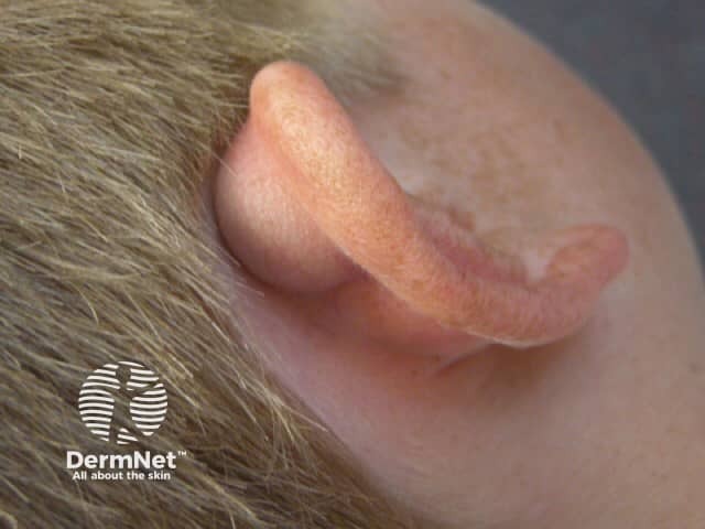
A cyst is a benign, round, dome-shaped encapsulatedlesion that contains fluid or semi-fluid material. It may be firm or fluctuant and often distends the overlying skin. There are several types of cyst. The most common are described here.
What is a pseudocyst?
Cysts that are not surrounded by a capsule are better known as pseudocysts. These commonly arise in acne.
Who gets cysts?
Cysts are very common, affecting at least 20% of adults. They may be present at birth or appear later in life. They arise in all races. Most types of cyst are more common in males than in females.
What causes cysts?
The cause of many cysts is unknown.
Epidermoid cysts are due to the proliferation of epidermal cells within the dermis. Their origin is the follicularinfundibulum. Multiple epidermoid cysts may indicate Gardner syndrome. The common term, sebaceous cyst, is a misnomer.
An epidermal inclusion cyst is a response to an injury. Skin is tucked in to form a sac that is lined by healthy epidermal cells that continue to multiply, mature and form keratin.
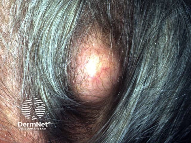
The origin of a trichilemmal cyst is hair root sheath. Inheritance is autosomal dominant (the affected gene is within the short arm of chromosome 3) or sporadic.
The origin of steatocystoma is the sebaceous duct within the hair follicle.Steatocystoma multiplex is sometimes an autosomal dominantly inherited disorder due to mutationslocalised to the keratin 17 (K17) gene, when it may be associated with pachyonychia congenita. More often, steatocysts are sporadic, when these mutations are not present.
The origin of the eruptive vellus hair cyst is follicular infundibulum. It may be inherited as an autosomal dominant disorder due to mutations in the keratin gene.
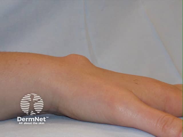
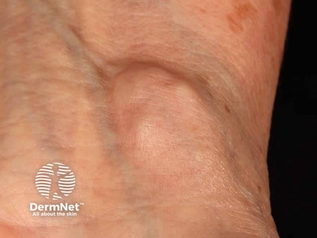
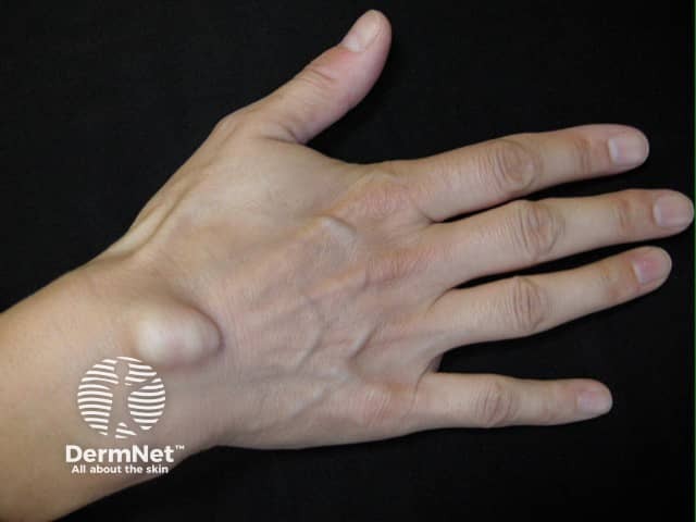
The origin of a ganglion cyst is degeneration of the mucoid connective tissue of a joint.
Occlusion of pilosebaceous units (hair follicles) or eccrine sweat ducts leads to a build-up of secretions, which can present as milia.
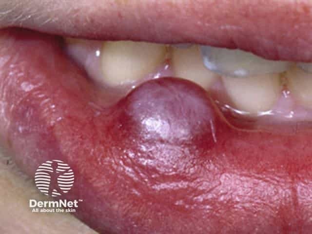
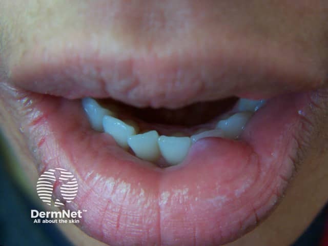
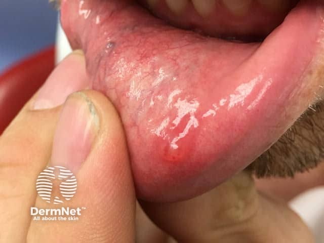
Occlusion of the orifice of a mucousgland can lead to a fluid-filled mucocoele in a mucous membrane (lip, vulva, vagina).
A milium is a pseudocyst due to failure to release keratin from an adnexal structure. The origin of primarymilium is infundibulum of the vellus hair follicle at the level of the sebaceous gland and is a miniature version of an epidermoid cyst. The source of secondary milium is a retention cyst within a vellus hair follicle, sebaceous duct, sweat duct or epidermis.
Pseudocysts in acne are formed by occlusion of the follicle by keratin and sebum.
What are the clinical features of cysts?
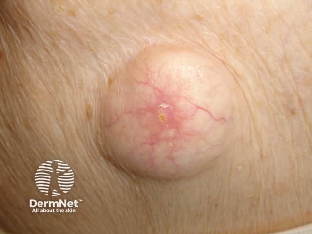
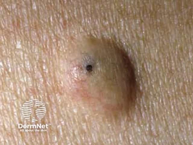
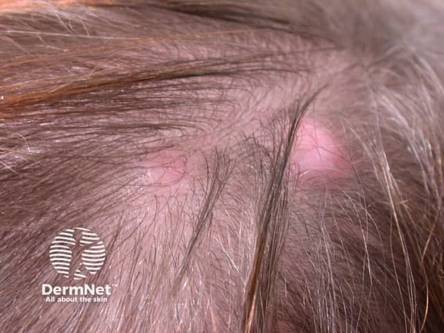
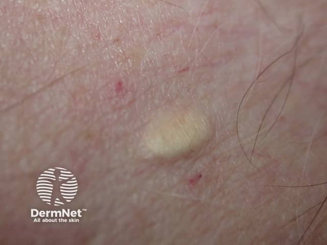
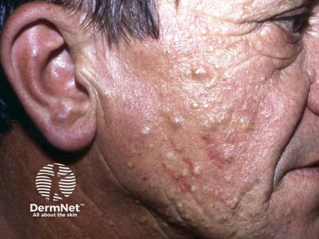
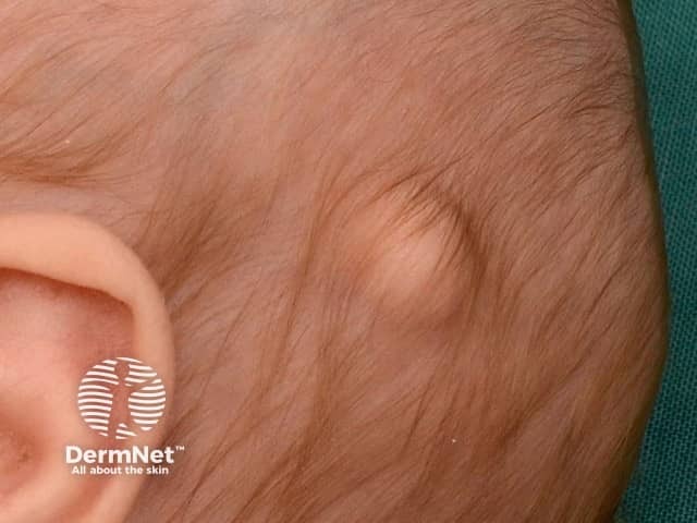
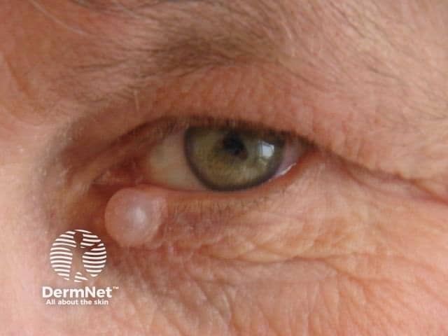
Epidermoid cyst
Epidermoid cysts occur on face, neck, trunk or anywhere where there is little hair.
Most epidermoid cysts arise in adult life.
They are more than twice as common in men as in women.
They present as one or more flesh–coloured to yellowish, adherent, firm, round nodules of variable size.
A central pore or punctum may be present.
Keratinous contents are soft, cheese-like and malodorous.
Scrotal and vulval cysts are frequently multiple and may calcify.
An epidermoid cyst is also called a follicular infundibular cyst, epidermal cyst, and keratin cyst.
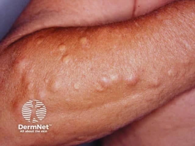
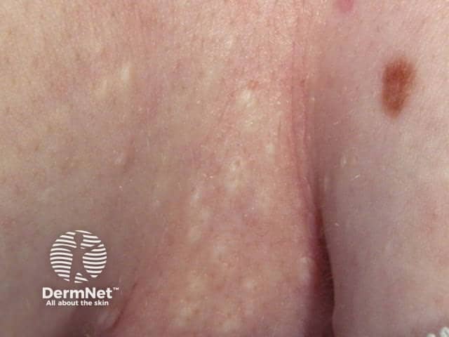
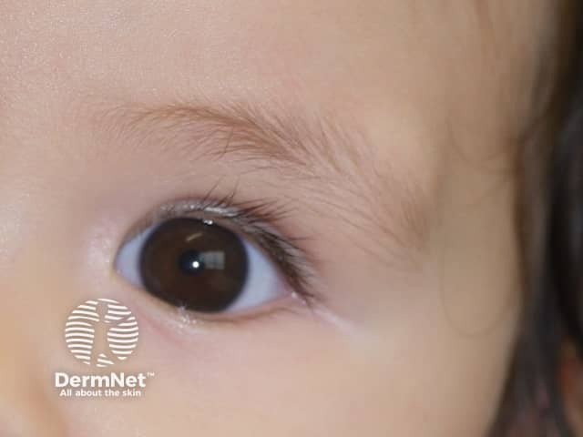
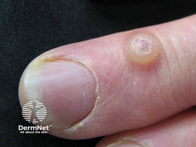
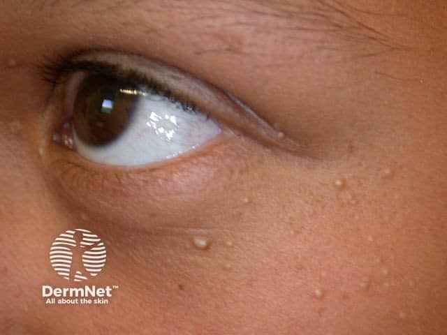
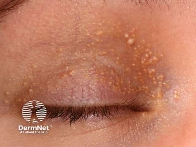
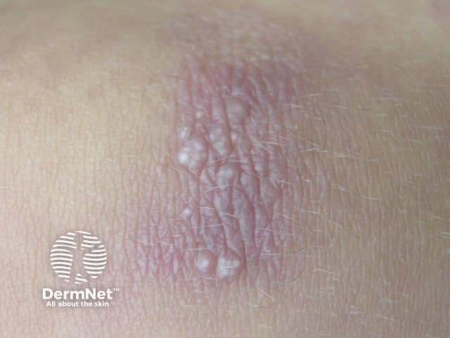
Milia are 1–2 mm superficial white dome-shaped papules containing keratin
Primary milia arise in neonates (50%), adolescents and adults; they are rarely familial and sometimes eruptive.
Primary milia occur on eyelids, cheeks, nose, gum margins (Bohn nodules) and palate (Epstein pearls) in babies; and eyelids, cheeks and nose of older children and adults.
Transverse primary milia are sometimes noted across the nasal groove or around the areola.
In milia en plaque, multiple milia arise on an erythematous plaque on face, chin or ears.
The contents of the cyst may penetrate the capsular wall and irritate the surrounding skin.
The area of tender, firm inflammation spreads beyond the encapsulated cyst.
Sterile pus may be discharged.
Secondary infection
A ruptured cyst may infrequently become secondarily infected by Staphylococcus aureus, forming a furuncle (boil).
Pressure effect
A dermoid cyst can cause pressure on underlying bony tissue.
A ganglion cyst can cause joint instability, weakness, limitation of motion and may compress a nerve.
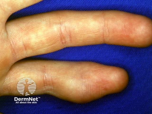
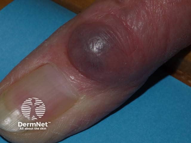
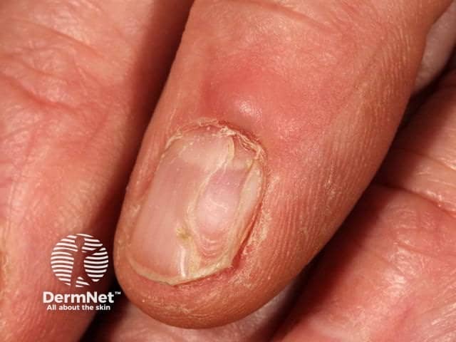
A digital mucous cyst may place pressure on the proximal matrix and cause a malformation of the nail.
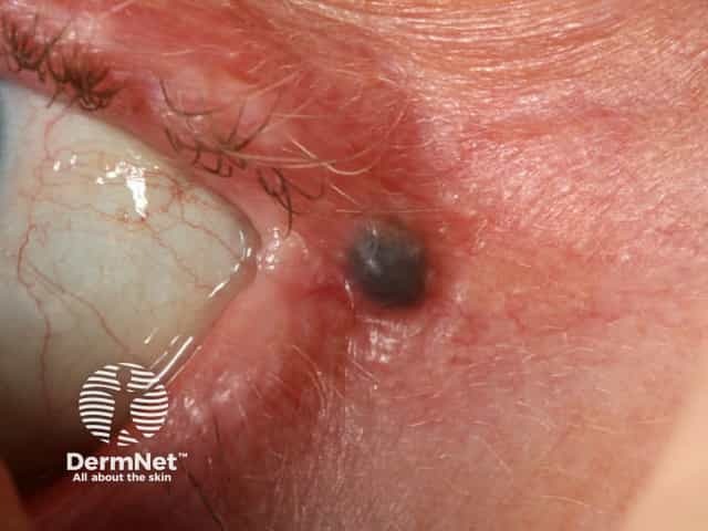
Malignancy
Cutaneous cysts and pseudocysts are non-proliferative benign lesions.
Nodulocystic basal cell carcinoma is a common skin cancer that presents as a rounded nodule and may initially be mistaken for a cyst, but steady enlargement, destruction of the epidermis with ulceration and bleeding occur eventually.
Cysts have typical clinical characteristics. When a cyst is surgically removed, it should undergo a histological examination. The type of lining of the wall of the cyst and the cyst contents help the pathologist classify it.
An epidermoid cyst is lined with stratified squamousepithelium that contains a granular layer. Laminated keratin contents are noted inside the cyst. An inflammatory response may be present in cysts that have ruptured.
Trichilemmal cysts have a palisaded outer layer without a granular layer. The contents are eosinophilic hair keratin. Older cysts may exhibit calcification. The proliferating variety is considered a tumour.
Steatocystoma has a folded cyst wall with prominent sebaceous gland lobules.
A dermoid cyst contains fully mature elements of the skin including fat, hairs, sebaceous glands, eccrine glands, and in 20%, apocrine glands.
The lining of the wall of a ganglion cyst or digital mucous cyst is collagen and fibrocytes. It contains hyaline material.
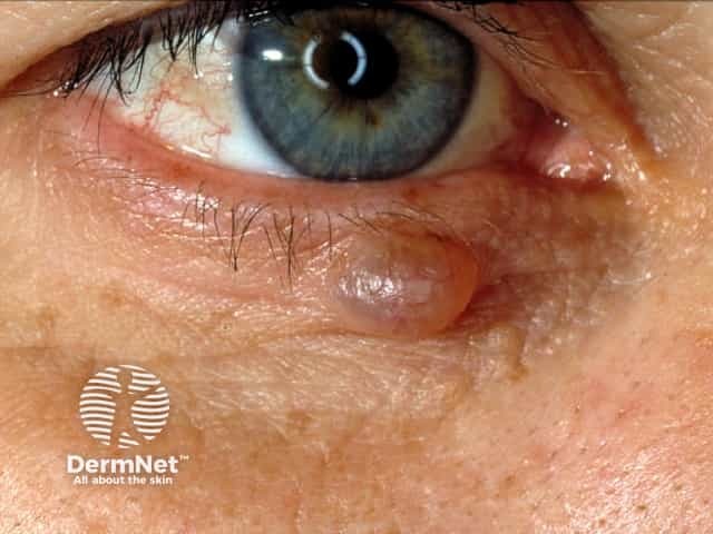
Hidrocystoma has a thin lining wall of eosinophilic bilaminar cells.
What is the treatment for cysts?
An asymptomaticepidermoid cyst does not need to be treated. In most cases, an attempt to remove only the contents of a cyst is followed by recurrence. If desired, cysts may be entirely excised. Recurrence is not uncommon, and re-excision may be surgically challenging.