Trichilemmal cyst is also known as pilar cyst. It is seen on the scalp and multiple cysts are common.
Histology of trichilemmal cyst
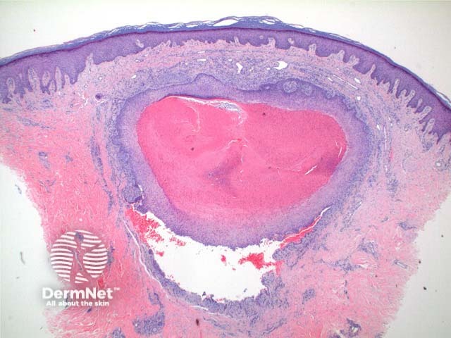
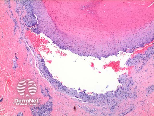
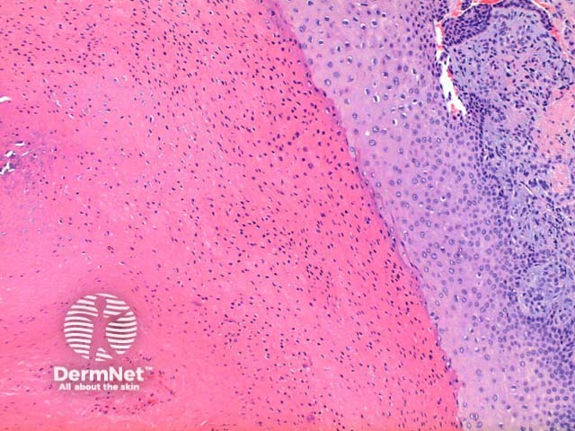
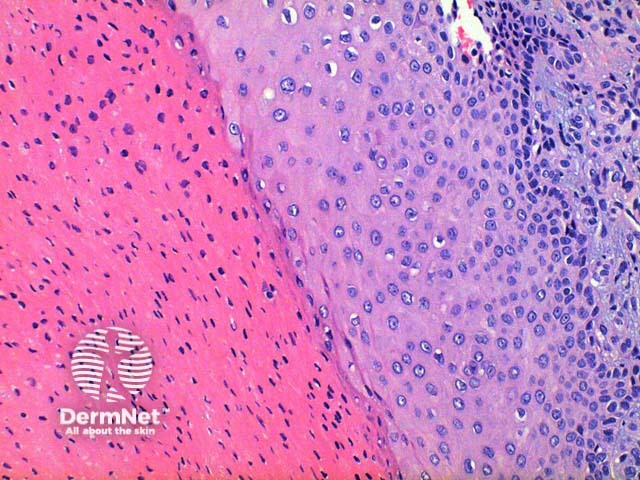
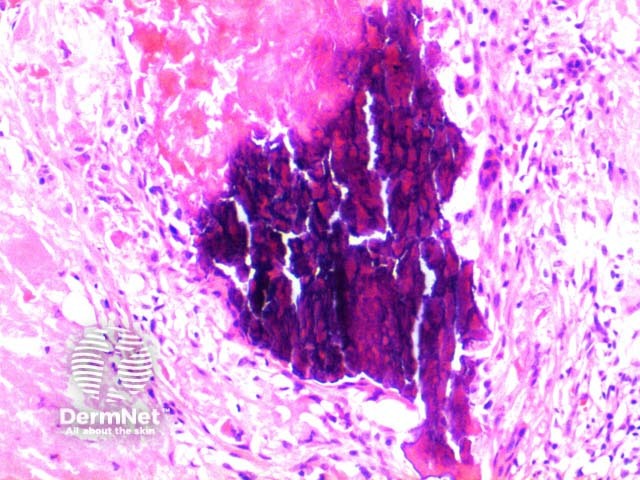
Scanning power view of trichilemmal cyst shows a epithelial lined cyst filled with brightly eosinophilic keratinaceous debris (Figure 1). Focal rupture of the cyst may occur with an associated giant cell reaction (Figure 2). Closer inspection of the cyst wall identifies trichilemmal differentiation (Figures 3 and 4) as occurs in the outer root sheath of the hairfollicle. This is seen as maturation of squamousepithelium with lack of a granular layer. The eosinophilic keratin centrally is densely packed frequently displaying cholesterolclefts. Focal calcification is seen in around 25% of cases (Figure 5).
Trichilemmal cyst pathology
Figure 1
Figure 2
Figure 3
Figure 4
Figure 5
Histological variants of trichilemmal cyst
Proliferating trichilemmal cyst: In this variant squamous proliferation can be seen arising from the cyst wall.
Malignant proliferating trichilemmal tumour: This tumour arises out of a pre-existing trichilemmal cyst. Clear transition is evident into an area of eccentric asymmetrical growth with malignant cytology
Differential diagnosis
Epidermalinclusion cyst: This cyst retains a granular layer within the maturing epithelial cyst wall.
Vellus hair cyst: The cyst contents contain multiple vellus hairs seen as small non pigmented hair shafts.
References
Skin Pathology (3rd edition, 2002). Weedon D
Pathology of the Skin (3rd edition, 2005). McKee PH, J. Calonje JE, Granter SR