
ADVERTISEMENT
You do not have any notes added to this page yet
Introduction
Demographics
Causes
Clinical features
Variation in skin types
Complications
Diagnosis
Treatment
Outlook
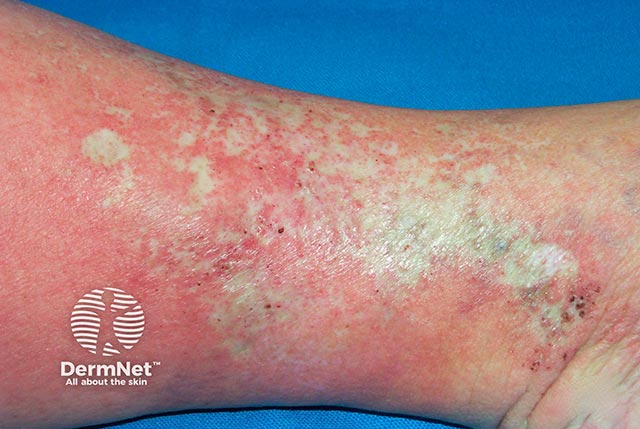
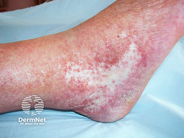
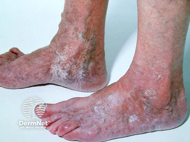
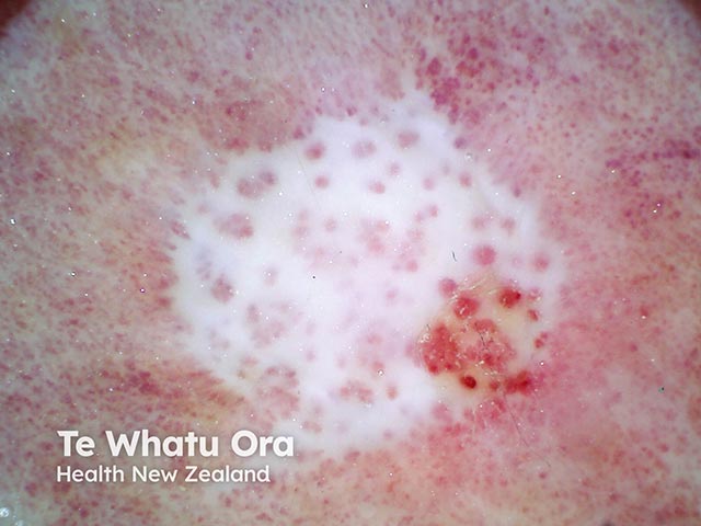
Atrophie blanche (white atrophy) is the name given to a particular type of scarring with ivory-white plaques accompanied by peripheral hyperpigmentation and telangiectasia. These usually occur on the lower extremities.
Atrophie blanche is a common feature of livedoid vasculopathy. The literature on this may be confusing as some people consider atrophie blanche to be a synonym for livedoid vasculopathy. However, atrophie blanche is more appropriately used to describe the morphologic pattern of scarring that occurs from healed ulcers.
For more images, click here
Atrophie blanche is an uncommon condition occurring primarily in middle-aged female adults. It is generally associated with patients with chronic venous insufficiency.
Atrophie blanche frequently occurs secondary to a condition called livedoid vasculopathy. Persistent painful ulcers as a result of livedoid vasculopathy leads to recurrent healing and scarring, causing atrophie blanche.
Atrophie blanche can also be due to other causes of recurrent ulceration in the lower limbs such as in:
Atrophie blanche can develop in sites of prior ulceration or in the absence of preceding ulcers.
The pathogenesis of atrophie blanche is thought to be secondary to the micro-occlusion of small blood vessels in the middle and deep dermis, which results in small tissue infarcts. This prevents the normal healing process. Blood vessel occlusion may be due to:
Atrophie blanche may be characterised by:
Scarring in atrophie blanche might appear different in patients with darker skin types.
Skin affected by atrophie blanche may be more susceptible to increased pain, ulceration, and infection.
Atrophie blanche is usually diagnosed with a good clinical history and examination.
Investigations should also be done to exclude any other underlying conditions like cutaneous small-vessel vasculitis, sickle cell disease, and antiphospholipid syndrome.
These investigations may include:
Treatment of atrophie blanche is directed to the underlying disease process that leads to this type of scarring. For example, in livedoid vasculopathy, drugs such as antiplatelets or anticoagulants are used to halt platelet aggregation and stimulate fibrinolysis.
The role of pentoxifylline may be considered in recurrent venous leg ulceration in those with peripheral vascular disease.
Compression therapy may speed up healing of wounds on the lower leg, particularly in venous disease. This can reduce the severity of atrophie blanche scar formation.
Other general measures include:
The prognosis of atrophie blanche is dependent on the underlying cause but it tends to be chronic and recurring.
An AI summary will appear based on your search term using data from all of the topic pages across the entire DermNet site.
Show more