Authors: Vanessa Ngan, Staff Writer, 2003. Revised: Dr Sashika Samaranayaka, Post Graduate Year 1, Department of Dermatology, Middlemore Hospital Auckland, New Zealand; Hon Assoc Prof Paul Jarrett, Dermatologist, Clinical Head Dermatology, Middlemore Hospital and Department of Medicine, The University of Auckland, Auckland, New Zealand. Copy edited by Gus Mitchell. January 2021.
Diabetic foot ulcer is a skin sore with full thickness skin loss on the foot due to neuropathic and/or vascular complications in patients with type 1 or type 2 diabetes mellitus.
Who gets diabetic foot ulcer?
Diabetic foot ulcer has an annual incidence of 2–6% and affects up to 34% of diabetic patients during their lifetime. Risk factors for developing a diabetic foot ulcer include:
Type 2 diabetes being more common than type 1
A duration of diabetes of at least 10 years
Poor diabetic control and high haemoglobin A1c
Being male
A past history of diabetic foot ulcer.
What causes diabetic foot ulcer?
Diabetic foot ulcers are caused by neuropathic and/or vascular complications of diabetes mellitus.
Neuropathic ulcer
High blood sugar levels can damage the sensory nerves resulting in a peripheralneuropathy, with altered or complete loss of sensation and an inability to feel pain. Peripheral neuropathy develops in approximately 50% of adults with diabetes, increasing the risk of injury to the feet from pressure, cuts, or bruises.
Vascular ulcer
Blood vessels can also be damaged by long-standing high blood sugar levels, decreasing blood flow to the feet (ischaemia) and/or skin (microangiopathy). This can result in poor wound healing.
What are the clinical features of diabetic foot ulcer?
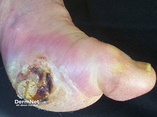
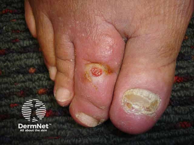
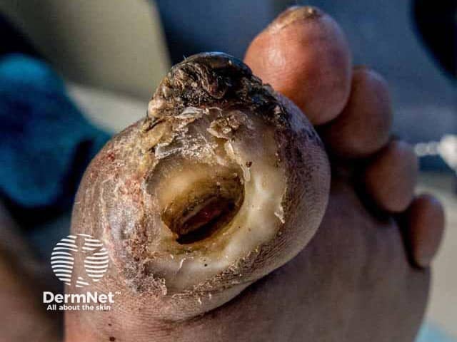
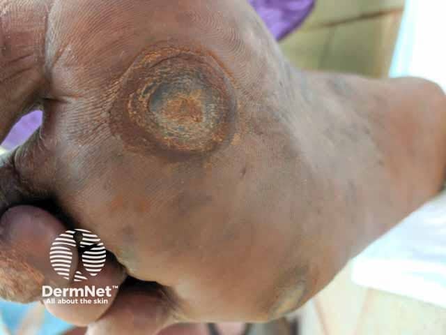
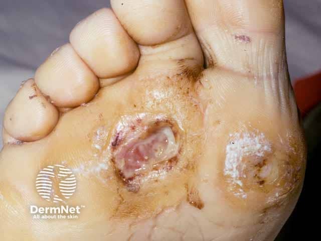
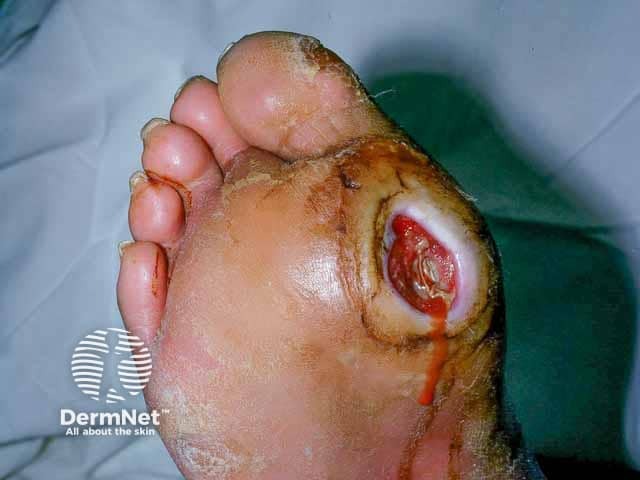
A diabetic foot ulcer is a skin sore with full thickness skin loss often preceded by a haemorrhagicsubepidermal blister. The ulcer typically develops within a callosity on a pressure site, with a circular punched out appearance. It is often painless, leading to a delay in presentation to a health professional. Tissue around the ulcer may become black, and gangrene may develop. Pedal pulses may be absent and reduced sensation can be demonstrated.
Foot ulcer at a pressure site
The severity of a diabetic foot ulcer can be graded and staged. There are many different classification systems. The University of Texas (UT) classification is a widely used, validated system (Table 1).
Table 1. University of Texas (UT) classification of diabetic foot ulcer
UT Grade
UT Stage
0: Pre- or post-ulcerative or healed wound
A: No infection or ischaemia
1: Superficial wound not involving tendon, capsule, or bone
B: Infection present
2: Wound penetrating to tendon or capsule
C: Ischaemia present
3: Wound penetrating to bone or joint
D: Infection and ischaemia present
UT Grade 1
UT Grade 2
UT Grade 3
What are the complications of diabetic foot ulcer?
Diabetic foot ulcer is particularly prone to secondary infection resulting in:
Wound infection
Cellulitis
Osteomyelitis.
How is diabetic foot ulcer diagnosed?
Diabetic foot ulcer is a clinical diagnosis of a painless foot ulcer in a patient with a long history of poorly controlled diabetes mellitus.
Investigations may include:
Swabs for secondary infection
X-rays for bone involvement
Angiography.
What is the differential diagnosis for diabetic foot ulcer?
An ulcer associated with other causes of peripheral neuropathy, such as leprosy
Optimise diabetes control to reduce neuropathic and vascular complications
Smoking cessation
Regular examination of the at-risk foot, and careful toenail trimming
Prompt treatment of non-ulcerative conditions such as tinea pedis or cracked heels
Appropriate footwear — properly fitting soft shoes or made-to-measure insoles
Exercise and physiotherapy
Education of patient, family, and healthcare providers
General measures
Multidisciplinary care — may include endocrinologist, podiatrist, diabetes education nurse, wound care nurse, neurologist, vascular surgeon, and/or orthopaedic surgeon
Adjuvant therapies — negative pressure wound therapy, ulcer excision and grafting, topical or hyperbaric oxygen, human growth factors, and/or amputation
What is the outcome for diabetic foot ulcer?
Diabetic foot ulcer may:
Heal over weeks to months
Re-ulcerate — recurrence rate is 40% within one year, 60% by three years, and 65% at five years
Result in lower extremity amputation in 14–24%.
The five-year mortality rate has been estimated to be 42%.
Bibliography
Armstrong DG, Boulton AJM, Bus SA. Diabetic foot ulcers and their recurrence. N Engl J Med. 2017;376(24):2367–75. doi:10.1056/NEJMra1615439. PubMed
Boulton AJM, Armstrong DG, Kirsner RS, et al. Diagnosis and management of diabetic foot complications. Arlington (VA): American Diabetes Association; October 2018. PubMed
Bourke J. Skin disorders in diabetes mellitus. In: Griffiths C, Barker J, Bleiker T, Chalmers R, Creamer D (eds) Rook’s Textbook of Dermatology, 9th edn. Wiley Blackwell, 2016: p64.2.
Everett E, Mathioudakis N. Update on management of diabetic foot ulcers. Ann N Y Acad Sci. 2018;1411(1):153–65. doi:10.1111/nyas.13569. PubMed
Hicks CW, Selvin E. Epidemiology of peripheral neuropathy and lower extremity disease in diabetes. Curr Diab Rep. 2019;19(10):86. doi:10.1007/s11892-019-1212-8. PubMed
Oyibo SO, Jude EB, Tarawneh I, Nguyen HC, Harkless LB, Boulton AJ. A comparison of two diabetic foot ulcer classification systems: the Wagner and the University of Texas wound classification systems. Diabetes Care. 2001;24(1):84–8. doi:10.2337/diacare.24.1.84. PubMed
Robinson TE, Kenealy T, Garrett M, Bramley D, Drury PL, Elley CR. Ethnicity and risk of lower limb amputation in people with Type 2 diabetes: a prospective cohort study. Diabet Med. 2016;33(1):55–61. doi:10.1111/dme.12807. PubMed
Zhang P, Lu J, Jing Y, Tang S, Zhu D, Bi Y. Global epidemiology of diabetic foot ulceration: a systematic review and meta-analysis. Ann Med. 2017;49(2):106–16. doi:10.1080/07853890.2016.1231932. PubMed