Authors: Dr Jenny Caesar, Dermatology Registrar, Singleton Hospital, Swansee, UK. November 2020; Updated: Dr Ian Coulson, Consultant Dermatologist, East Lancashire NHS Trust, UK. Copy edited by Gus Mitchell. August 2021
Venous insufficiency occurs when the normal flow of blood from the superficial veins to the heart via the perforating deep veins in the lower limbs is impaired, resulting in chronic venous congestion. It can be classified as superficial vein insufficiency, perforating, or deep vein insufficiency.
Who gets venous insufficiency?
Venous insufficiency is common, affecting all races and both sexes. Estimates suggest rates as high as 50% in some populations. A US study found ethnic whites had a higher rate of venous insufficiency compared to Hispanics, African Americans, and Asians. The Edinburgh Vein Study reported the incidence of chronic venous insufficiency was similar in men and women. Prevalence increases with age, obesity, a family history of varicose veins, and multiple pregnancies.
What causes venous insufficiency?
The venous system in the lower legs consists of a low-pressure superficial network connected by perforating veins to a high-pressure deep network. Venous blood flows from the superficial to deep veins by the action of the calf muscle pump. Retrograde flow is prevented by competent valves.
Chronic venous insufficiency can be caused by:
Venous factors
Incompetent valve in the superficial, perforating, and deep systems
External compression of veins
Deep venous obstruction
Previous deep vein thrombosis
Impaired calf muscle pump
Immobility including lack of physical exercise and paralysis
Once venous hypertension is established, the cutaneous features of chronic venous insufficiency are caused by:
Leukocytes trapped in capillaries release proteolyticenzymes and oxygen free radicals which damage the capillarybasement membrane
Plasmaproteins, such as fibrinogen, leak into the surrounding tissues, forming a fibrin cuff which reduces oxygen delivery to the tissues
Inflammation and tissue loss.
What are the clinical features of venous insufficiency?
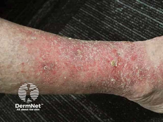
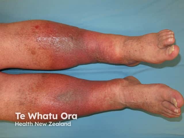
Superficial venous insufficiency can be asymptomatic but may cause aching, cramping, throbbing, burning, or heaviness in the leg. Pain is typical of deep venous insufficiency. Symptoms improve with leg elevation.
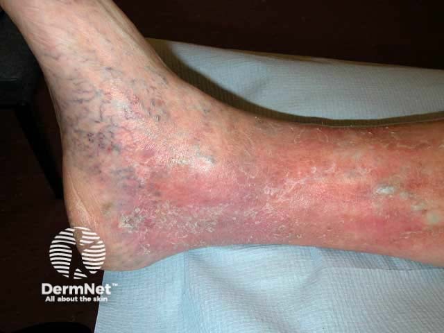
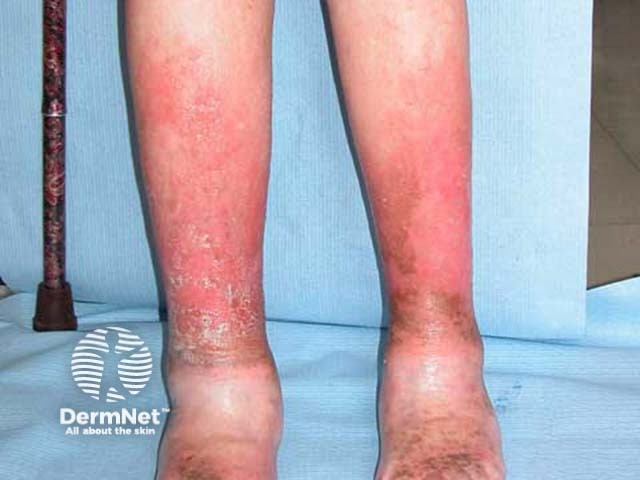
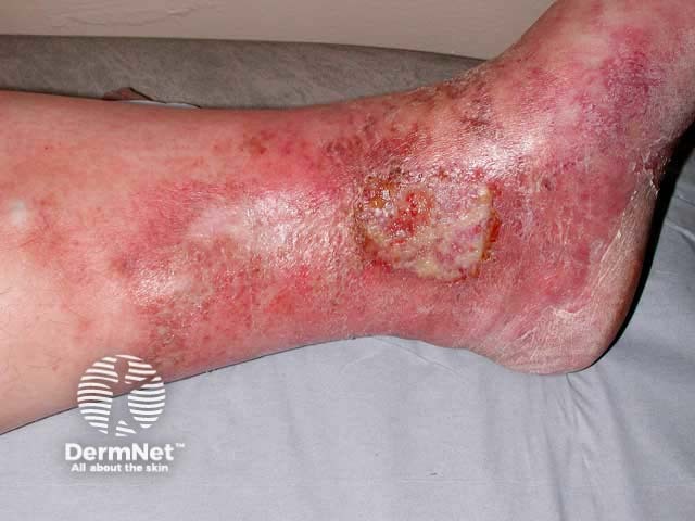
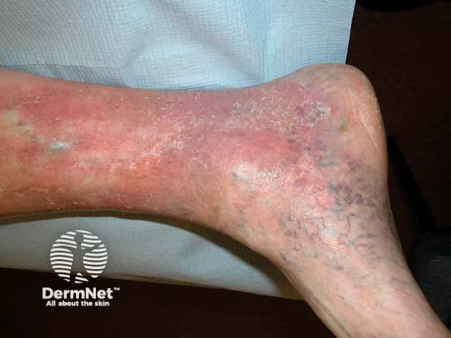
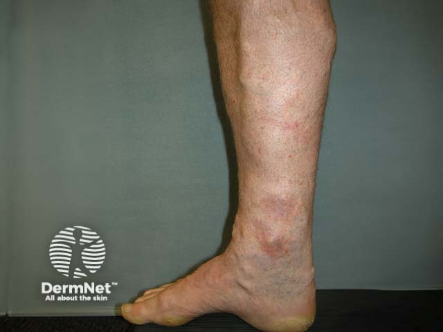
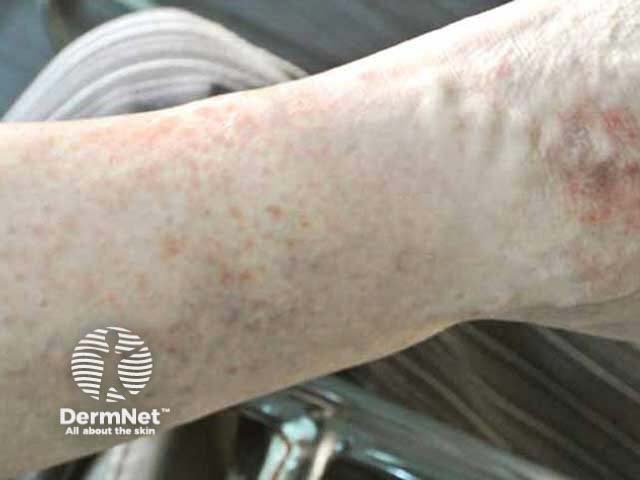
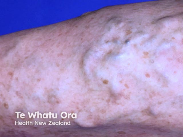
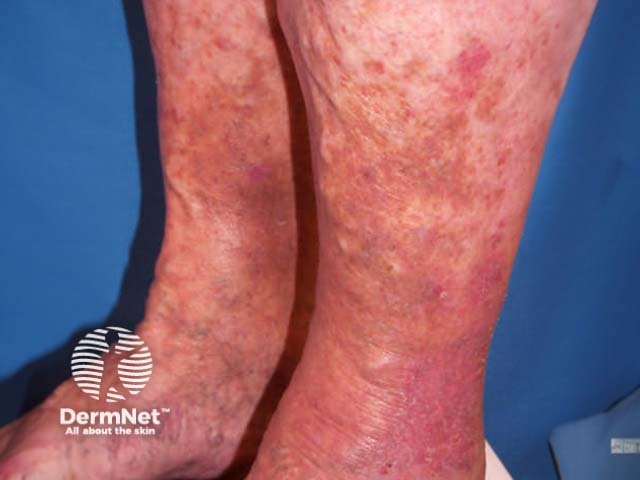
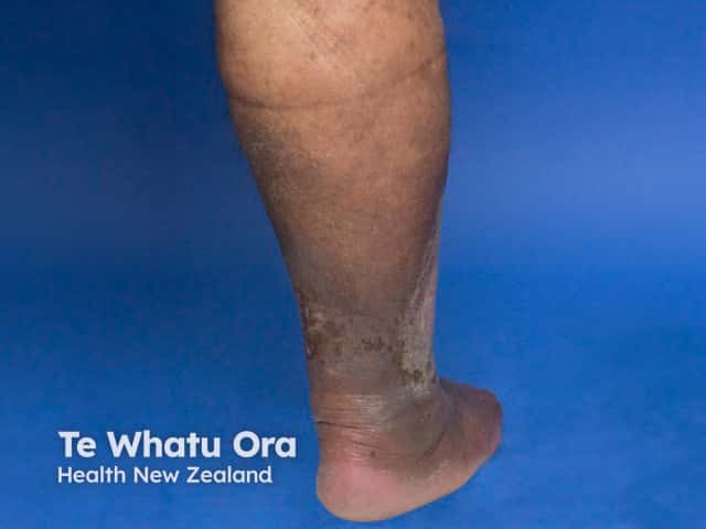
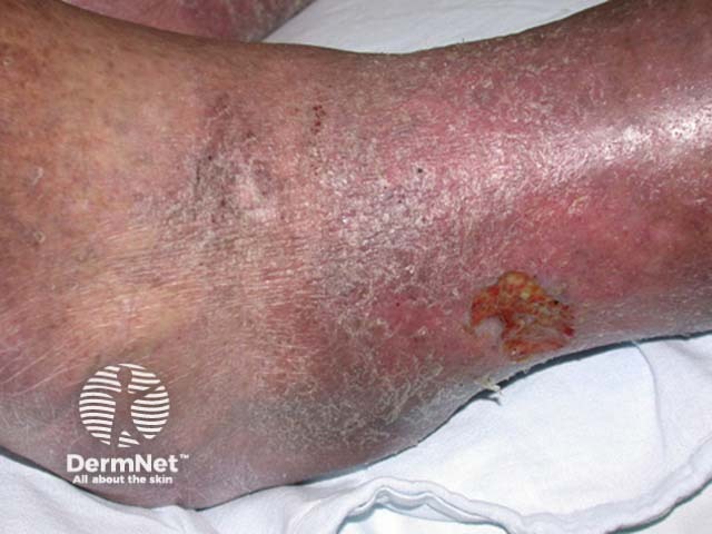
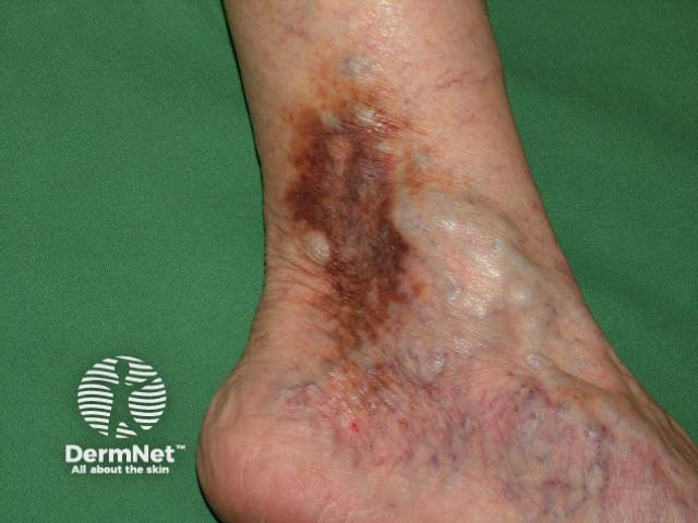
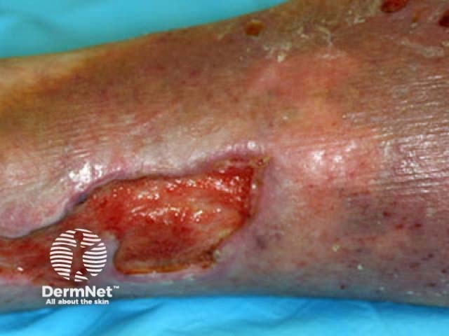
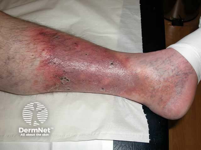
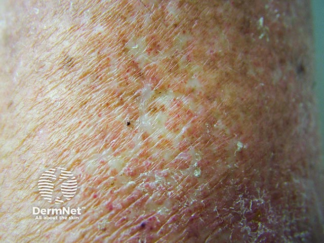
Clinical signs of chronic venous insufficiency include:
Pitting oedema around the ankle, worse at the end of the day
Corona phlebectatica paraplantaris (malleolar flare) on the inner aspect of the ankle and instep – fan-shaped distribution of blue and red telangiectases (intradermal and superficial venules), capillary stasis spots (red-purple coin-shaped areas formed by papillary capillaries), and venous cups (dilated converging veins on the instep)
Pigmentation can be pinpoint or a patchy rusty-red due to extravasation of red blood cells and haemosiderin accumulation in the dermis
Varicosities.
Clinical features of venous insufficiency
End-of-day ankle oedema
Corona phlebectatica and venous eczema
Varicosities and venous eczema
Pigment changes in venous insufficiency
Pinpoint pigmentation
Varicosities and pigmentation
Patchy rusty-red pigmentation
How do clinical features vary in differing types of skin?
Venous insufficiency in Thai patients has been reported to be less likely to be associated with pain, oedema, inflammation, and visible varicose veins compared to ethnic whites.
What are the complications of venous insufficiency?
Venous insufficiency is usually diagnosed on history and examination. Varicose veins may require the patient to be standing to appreciate. Duplex ultrasonography is the preferred investigation to demonstrate reflux and communications between the deep and superficial venous networks.
The CEAP (Clinical, Etiologic, Anatomic, Pathologic) system is used categorise and classify venous insufficiency but is unhelpful as a severity score. [see details Varicose veins]
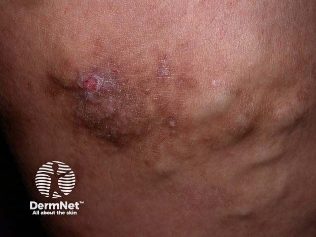
The corona phlebectatica paraplantaris is a clinical sign of severe venous stasis. Specifically, blue telangiectases and capillary stasis spots may be the most useful correlates with the severity of chronic venous insufficiency.
What is the differential diagnosis for venous insufficiency?
Varicose veins — sclerotherapy, endovenous laser therapy, radiofrequency ablation, surgery [see Leg vein therapies]
What is the outcome for venous insufficiency?
Venous insufficiency is inexorably progressive. The rate of complication development is not known.
Most patients with chronic venous insufficiency will experience at least intermittent episodes of venous eczema.
Leg ulceration is the most important complication and is a major burden for health systems throughout the world. Estimates suggest 4% of patients with varicose veins will develop venous leg ulcers in their lifetime.
Bibliography
Chiesa R, Marone EM, Limoni C, Volontè M, Petrini O. Chronic venous disorders: correlation between visible signs, symptoms, and presence of functional disease. J Vasc Surg. 2007;46(2):322–36 doi:10.1016/j.jvs.2007.04.030. Journal
Criqui MH, Jamosmos M, Fronek A, et al. Chronic venous disease in an ethnically diverse population: the San Diego Population Study. Am J Epidemiol. 2003;158(5):448–56. doi:10.1093/aje/kwg166. Journal
Jawien A. The influence of environmental factors in chronic venous insufficiency. Angiology. 2003;54 Suppl 1:S19–S31. doi:10.1177/0003319703054001S04. PubMed
Labropoulos N. How does chronic venous disease progress from the first symptoms to the advanced stages? A Review. Adv Ther. 2019;36(Suppl 1):13–9. doi:10.1007/s12325-019-0885-3. Journal
Langer RD, Ho E, Denenberg JO, Fronek A, Allison M, Criqui MH. Relationships between symptoms and venous disease: the San Diego population study. Arch Intern Med. 2005;165(12):1420–4. doi:10.1001/archinte.165.12.1420. PubMed
Necas M. Duplex ultrasound in the assessment of lower extremity venous insufficiency. Australas J Ultrasound Med. 2010;13(4):37–45. doi:10.1002/j.2205-0140.2010.tb00178.x. PubMed Central
Novak CJ, Khimani N, Kaye AD, Jason Yong R, Urman RD. Current therapeutic interventions in lower extremity venous insufficiency: a comprehensive review. Curr Pain Headache Rep. 2019;23(3):16. doi:10.1007/s11916-019-0759-z. PubMed
Robertson L, Lee AJ, Evans CJ, et al. Incidence of chronic venous disease in the Edinburgh Vein Study. J Vasc Surg Venous Lymphat Disord. 2013;1(1):59–67. doi:10.1016/j.jvsv.2012.05.006. PubMed
Uhl JF, Cornu-Thenard A, Satger B, Carpentier PH. Clinical analysis of the corona phlebectatica. J Vasc Surg. 2012;55(1):150–3. doi:10.1016/j.jvs.2011.04.070. Journal
Youn YJ, Lee J. Chronic venous insufficiency and varicose veins of the lower extremities. Korean J Intern Med. 2019;34(2):269–83. doi:10.3904/kjim.2018.230. Journal