Pitted keratolysis, also known as keratolysis plantare sulcatum or ringed keratolysis, is a superficial bacterial skin infection characterised by crater-like pits and malodour. It typically affects pressure-bearing areas on the soles of the feet, although the palms are rarely affected.
This condition is very treatable with a good prognosis.
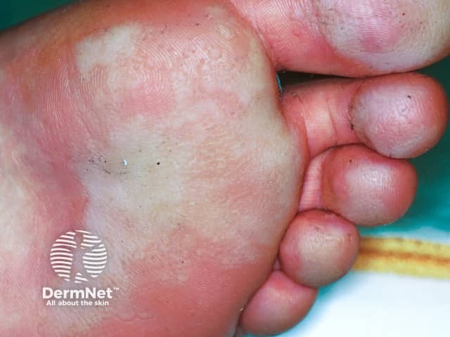
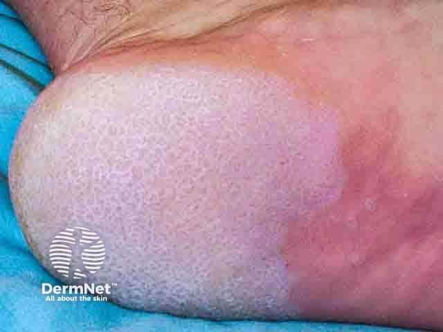
Pits and maceration in pitted keratolysis on the heel (PK-patient1)
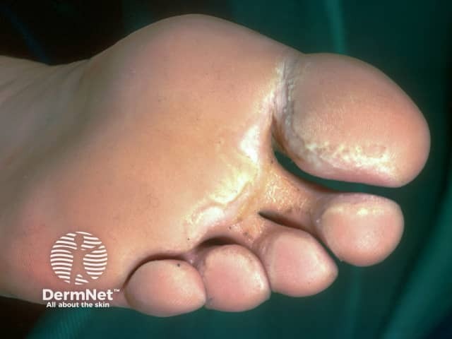
Interdigital and toe pitted keratolysis
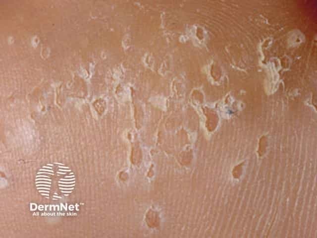
Close up of plantar pits in pitted keratolysis
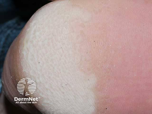
White macerated and pitting plantar skin in pitted keratolysis
Pitted keratolysis is caused by a range of bacterial species. The most common are Corynebacteria, Dermatophilus congolensis, Kytococcus sedentarius, Actinomyces, or Streptomyces.
Bacteria thrive under moist and warm conditions. They proliferate and produce proteaseenzymes that cause destruction of the stratum corneum to create pits/craters. The odour is associated with the sulfur compounds (thiols, sulphides, and thioesters) which are produced by the bacteria.
What are the clinical features of pitted keratolysis?
The infection often occurs bilaterally on pressure-bearing areas, most commonly the ball of the foot and heel. Involvement of the palms have been reported in certain professions such as rice paddy farmers.
It is often asymptomatic, however, when symptomatic there may be associated pruritus and pain on walking.
Characteristic features include:
Pits on the stratum corneum (1–3mm); these may form confluences, irregular erosions, or sulci
Some pits may have a brown appearance (giving the appearance of dirty feet)
The appearance of the pits are often amplified when feet are wet.
How do clinical features vary in differing types of skin?
Pitted keratolysis has been seen in patients of all skin types. They all present similarly and can present in a range of sizes and colours.
What are the complications of pitted keratolysis?
Psychosocial impact due to foot odour
Limitation of function due to symptomatic pitted keratolysis
No mortality is associated with pitted keratolysis.
How is pitted keratolysis diagnosed?
Diagnosis is often clinical given its distinctive appearance and malodour. Consider examination of intertriginous areas (axilla and groin) for co-existing corynebacterial infections, such as erythrasma and trichomycosis axillaris.
de Almeida HL Jr, Siqueira RN, Meireles Rda S, Rampon G, de Castro LA, Silva RM. Pitted keratolysis. An Bras Dermatol. 2016;91(1):106–8. doi:10.1590/abd1806-4841.20164096. Journal
Kaptanoglu AF, Yuksel O, Ozyurt S. Plantar pitted keratolysis: a study from non-risk groups. Dermatol Reports. 2012;4(1):e4. Published 2012 Feb 7. doi:10.4081/dr.2012.e4. Journal
Ramsey ML. Pitted keratolysis: a common infection of active feet. Phys Sportsmed. 1996;24(10):51–6. doi:10.3810/psm.1996.10.1319. Journal
Shelley WB, Shelley ED. Coexistent erythrasma, trichomycosis axillaris, and pitted keratolysis: an overlooked corynebacterial triad?. J Am Acad Dermatol. 1982;7(6):752–7. doi:10.1016/s0190-9622(82)80158-8. Journal
Vlahovic TC, Dunn SP, Kemp K. The use of a clindamycin 1%-benzoyl peroxide 5% topical gel in the treatment of pitted keratolysis: a novel therapy. Adv Skin Wound Care. 2009;22(12):564–6. doi:10.1097/01.ASW.0000363468.18117.fe. Journal