
ADVERTISEMENT
You do not have any notes added to this page yet
Introduction
Demographics
More information
Clinical features
Complications
Diagnosis
Differential diagnoses
Treatment
Outcome
Epstein-Barr virus is one of the most ubiquitous human viruses, with up to 95% of adults worldwide having been infected.
Epstein-Barr virus (EBV) is also called Human herpesvirus 4 (HHV-4).
Epstein-Barr virus is a member of the order Herpesvirales, family Herpesviridae, subfamily Gammaherpesvirinae, genus Lymphocriptovirus.
Herpes viruses are characterised by:
Epstein-Barr virus may be passed from person to person via:
The incubation period is about six weeks. Following primary infection, EBV enters a latent phase in B lymphocytes from which it can be reactivated, especially if immunocompromised. Virus shedding and transmission can occur during primary infection or intermittently during reactivation.
Epstein-Barr virus is best known for causing infectious mononucleosis (glandular fever) in adolescents and young adults, although primary EBV infection can be asymptomatic (10%) particularly in children.
EBV can also cause other disorders with mucocutaneous features, and has been implicated in the pathogenesis of many more.
Disorder |
Mucocutaneous features |
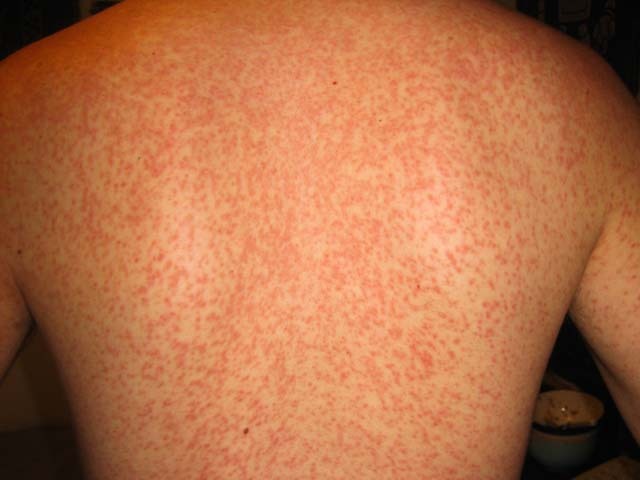
Infectious mononucleosis |
Palatal petechiae, rash, eyelid/periorbital swelling |
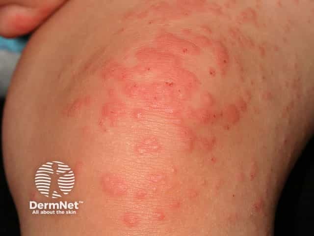
Papular rash over buttocks, thighs, arms, face |
|
Painful deep punched out ulcer(s) on the genitalia |
|
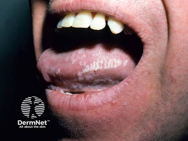
Asymptomatic white plaques on the lateral tongue |
|
Symmetrical painful erythema and oedema of hands and feet |
|
Pink papules beginning in the armpit or groin, spreading along one side of the trunk |
|
Photosensitivity |
|
One form is classified as chronic active EBV infection in childhood |
|
Chronic active EBV |
Persistent or recurrent IM-like symptoms including of the skin unable to be explained by other known processes |
Jaundice, itch |
EBV, Epstein-Barr virus; IM, infectious mononucleosis
EBV has been postulated to trigger a number of autoimmune conditions in genetically predisposed individuals: examples include systemic lupus erythematosus, Sjögren syndrome, and dermatomyositis.
Disorder |
Role of EBV |
Both the acute and chronic forms have been postulated to be triggered by infectious agents |
|
EBV DNA has been detected by PCR in some (but not all) tissue samples of LCH |
|
Congenital infection by EBV can present with this phenotype |
EBV, Epstein-Barr virus; LCH, Langerhans cell histiocytosis; PCR, polymerase chain reaction
Epstein-Barr virus is implicated in many lymphoproliferative disorders and tumours. Examples include:
Epstein-Barr virus infection will be considered on the clinical presentation and confirmed on investigations which may include:
[see Laboratory tests for viral infections]
Epstein-Barr virus infection becomes latent with the risk of reactivation in later life.
The age at which EBV is acquired influences the clinical picture and complications. Primary infection in adolescents and adults is more likely to result in infectious mononucleosis than if infection occurs in childhood, and IM carries an increased risk of subsequent development of Hodgkin lymphoma. Primary infection in early childhood is associated with subsequent nasopharyngeal carcinoma or Burkitt lymphoma.
An AI summary will appear based on your search term using data from all of the topic pages across the entire DermNet site.
Show more