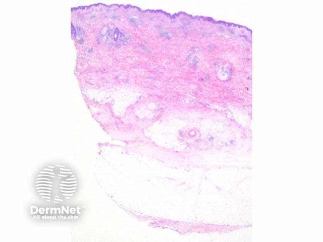
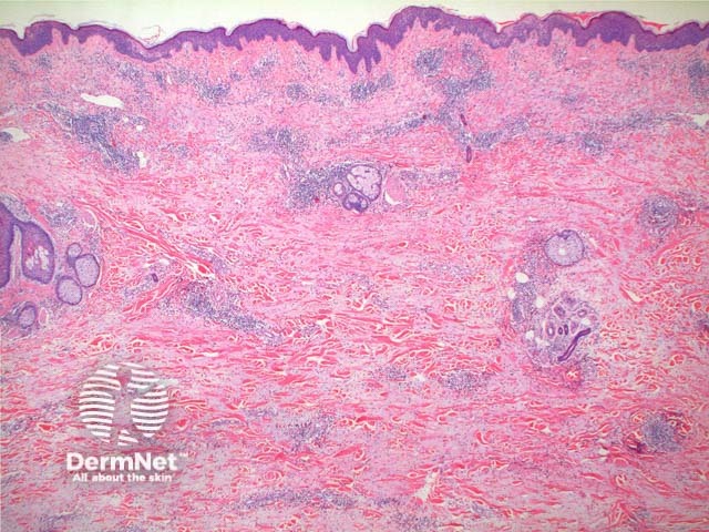
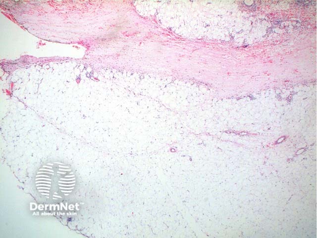
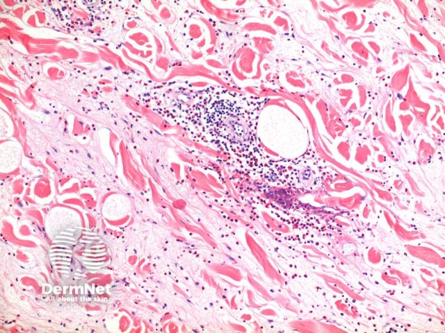
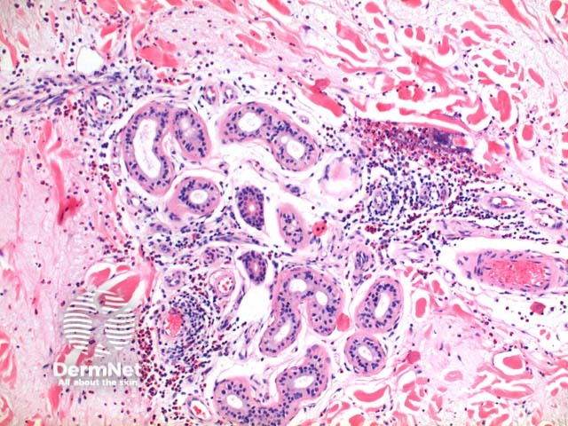
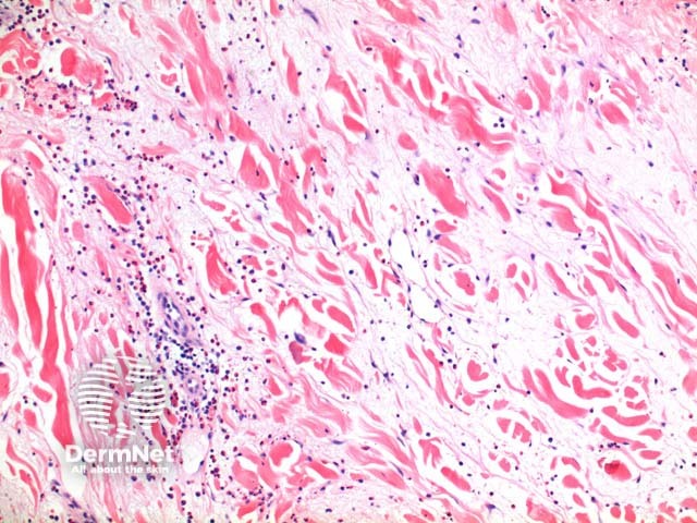
Scanning power view of Wells syndrome reveals a superficial and deep perivascular and interstitialinflammatory pattern (Figures 1 and 2). This can be seen to extend into the subcutaneous tissue (Figure 3) or even the underlying muscle. The inflammatory infiltrate is comprised of lymphocytes, histiocytes and abundant eosinophils (Figures 4,5 and 6). Degranulation of the eosinophils is seen forming flame figures (Figures 4 and 5). In this particular case extensive interstitial mucin is seen (Figures 4 and 5).
Figure 1
Figure 2
Figure 3
Figure 4
Figure 5
Figure 6
Variants of Wells syndrome pathology
Bullous Wells disease: A subepidermal blister can form in the presence of prominent papillary dermaloedema.
Differential diagnosis of Wells syndrome pathology
Insect bite reaction: Typically here the infiltrate is more localised forming a wedge-shaped pattern possibly with focal overlying epidermal changes. In some cases discrimination is not possible and clinical correlation is required.
Bullous pemphigoid: In most circumstances, it is the urticarial phase of bullous pemphigoid which may prove difficult to discriminate. Eosinophils can be seen to tag along with the dermoepidermal junction in conjunction with basal layervacuolar degeneration
Churg Strauss syndrome: While dermal eosinophilia and flame figures can be seen, this condition is characterised by necrotisinggranuloma formation and variable degrees of vasculitis.
References
Skin Pathology (2nd edition, 2002). Weedon D
Pathology of the Skin (3rd edition, 2005). McKee PH, J. Calonje JE, Granter SR
Churg, A. Recent advances in the diagnosis of Churg-Strauss syndrome. Mod Pathol 2001;14(12):1284–93. PubMed