Reticulohistiocytoma (reticulohistiocytosis) is a non-Langerhans cell histiocytosis. Lesions may be solitary (for example solitary cutaneous reticulohistocytoma) or multiple (for example multicentric reticulohistiocytosis). Multicentric reticulohistocytosis is characterized by cutaneous or mucosalpapular lesions associated with severe polyarthritis and arthralgias.
Histology of reticulohistiocytoma
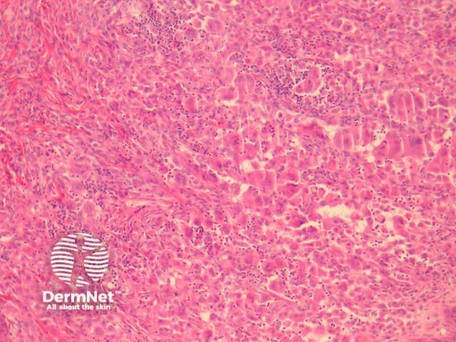
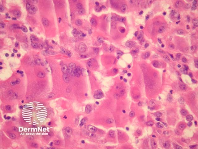
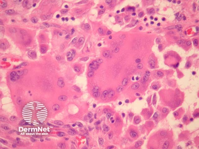
In reticulohistiocytoma, sections show a diffuseinfiltration of numerous large, mononucleated or multinucleatedhistiocytes in the dermis (figures 1-3). There are associated lymphocytes and dermalfibrosis. Characteristically, the lesional cells have a dense pink cytoplasm, variously referred to as “oncocytic” or “ground glass” (figures 2, 3).
Figure 1
Figure 2
Figure 3
Special studies for reticulohistiocytoma
Reticulohistiocytoma cells are positive with CD68 and Factor 13a. S100 and CD1a are typically negative.
Differential diagnosis of reticulohistiocytoma
Other histiocytic diseases: these typically lack the characteristic “ground glass” cytoplasm seen in reticulohistiocytoma. In addition to morphologic features, CD1a and S100 immunohistochemical studies can be used to exclude Langerhans cellhistiocytosis and Rosai-Dorfman disease respectively.
Juvenile xanthogranuloma shows “Touton” giant cells, and lipidisation of histiocytes. The rarer histocytoses may require more extensive immunohistochemical studies and clinical correlation.
References
Jung HD, Kim HS, Lee JY, Kim HO, Park YM. Multicentric reticulohistiocytosis. Acta Derm Venereol. 2013 Jan;93(1):124–5.