
ADVERTISEMENT
You do not have any notes added to this page yet
Introduction
Demographics
Causes
Symptoms
Clinical signs
Diagnosis
Treatment
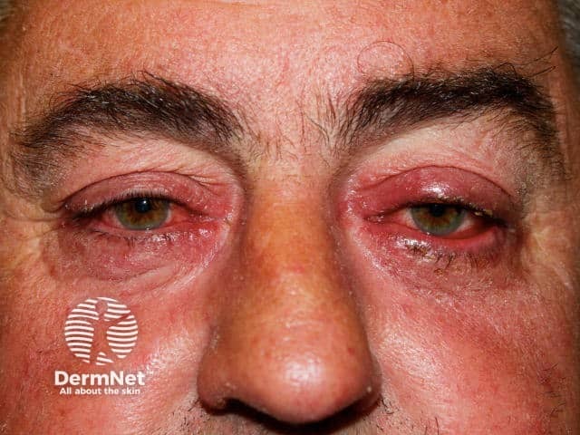
Rosacea is a common skin problem in which there is mid-facial flushing, redness, prominent vasculature, swelling, papules and/or pustules. Ocular rosacea is a form of rosacea that involves the eyelids and the front of the eye. Ocular rosacea includes:
Ocular rosacea affects adult males and females equally, with one study reporting an average age at presentation of 56 years. It is uncommon in children and generally starts after the age of 30 years.
It usually occurs in patients with existing rosacea but it can be the first sign of the disease. Ocular rosacea tends to occur in patients with facial flushing (1).
The exact cause of ocular rosacea is unknown. However, immunological factors, micro-organisms on the skin surface, and reactive blood vessels are involved (1).
Obstruction of meibomian glands changes tear film composition leading to:
Ocular rosacea mostly affect the eyelids, conjunctiva and cornea. Rarely, it can involve the iris and sclera. Symptoms can include:
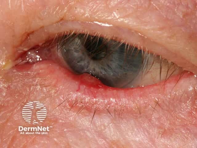
Anterior blepharitis involves the lid margin and lash line. Signs include:
Posterior blepharitis involves obstruction of the ducts and loss of the Meibomian glands (2). This leads to:
An inflamed cornea (keratitis) is a rare but serious ocular complication of rosacea and can threaten vision (1).
Iritis, episcleritis and scleritis are rare in ocular rosacea. They cause a painful, watery red eye and may affect vision.
Ocular rosacea may be suspected in a patient with cutaneous rosacea that has eyelid or eye disease. The symptoms and signs are nonspecific, so the diagnosis is more difficult in the absence of cutaneous rosacea.
Blepharitis can also be due to seborrhoeic dermatitis, a scaly skin condition, and inflammatory papules on and around the eyelids may be due to periorificial dermatitis.
Anterior blepharitis can be successfully treated with various topical antiseptics and antibiotics including:
The following oral antibiotics are used for ocular rosacea:
They reduce bacteria, improve tear film stability and normalise meibonian gland secretions.
Oral antibiotics are generally continued for 6–12 weeks, and then slowly tapered over the course of one to two months. Further courses of oral antibiotics can be used for disease flare-ups.
Styes failing to clear with topical antibiotic are treated with oral anti-staphylococcal antibiotics such as flucloxacillin.
Oral isoretinoin can be used in low dose to treat ocular rosacea but with caution because its adverse effects include increased infections, dry eye, and other ocular effects.
Oral omega-3 fatty acid supplementation has been reported to be beneficial for some patients with dry eyes.
Surgery may be required to repair corneal opacification or perforation due to rosacea keratitis. The procedure is called keratoplasty.
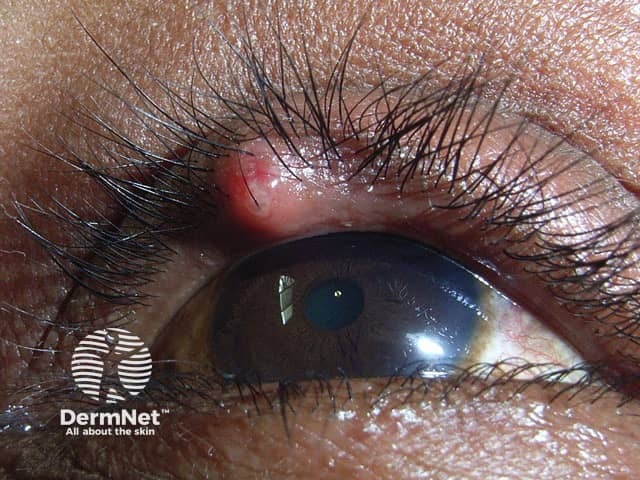
Hordeola that fail to improve with warm compresses and antibiotic therapy may be excised.
An AI summary will appear based on your search term using data from all of the topic pages across the entire DermNet site.
Show more