Obesity-associated lymphoedematous mucinosis — extra information
Extra information
ADVERTISEMENT
Obesity-associated lymphoedematous mucinosis
Last reviewed: December 2023
Author(s): Dr Alpana Mohta, Consultant Dermatologist, Sharda Multispeciality Clinic, India; Dr Libby Whittaker, DermNet Medical Writer, New Zealand (2023)
Reviewing dermatologist: Dr Ian Coulson
Edited by the DermNet content department
What is obesity-associated lymphoedematous mucinosis?
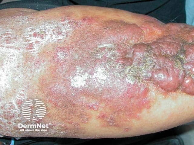
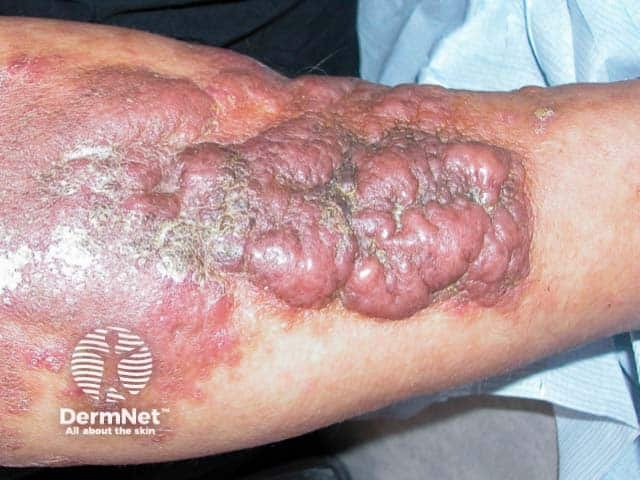
Obesity-associated lymphoedematous mucinosis (OALM) is a rare and recently identified form of pretibialdermal mucinosis that affects individuals who are obese. It displays clinical similarities to pretibial myxoedema but occurs in patients without thyroid dysfunction.
OALM has been previously referred to as chronic obesity lymphoedematous mucinosis.
Floridplaques of obesity-associated lymphoedematous mucinosis on the shins (OALM-patient1)
Florid plaques of obesity-associated lymphoedematous mucinosis on the shins (OALM-patient1)
Euthyroid pretibial mucinosis, the spectrum of which includes OALM, may also be associated with venous insufficiency, lymphoedema, and trauma.
What causes obesity-associated lymphoedematous mucinosis?
Cutaneous mucinoses involve aberrantdeposition of mucin, sulfated glycosaminoglycans (GAG), and hyaluronic acid in the dermis.
The specific underlying pathomechanism of OALM is partially understood.
In obese patients, lymphaticstasis can lead to local oxygen deficiency.
Obesity-associated tissue hypoxia and venous insufficiency cause the accumulation of plasmaproteins like albumin, fibrinogen, and coagulation factors in the interstitial space.
Interstitial oedema further diminishes local oxygen supply. Consequently, fibroblasts respond by increasing the synthesis and deposition of glycosaminoglycans and mucin, which leads to the formation of dermal mucinosis.
What are the clinical features of obesity-associated lymphoedematous mucinosis?
The clinical signs of OALM include asymptomaticpapules and nodules that develop into plaques.
Typically these are skin-coloured to erythematous or yellowish in colour.
The shins are most commonly affected.
The lesions are accompanied by pitting oedema and lymphostasis.
Uncommonly, subepidermal blisters form due to dermal oedema.
With time, lymphoedema develops in the lower limbs, usually bilateral and sparing the feet and ankles. In severe cases, vesicles may develop.
OALM may be seen in conjunction with stasis mucinosis, which is characterised by blue-violet, partially blanchable plaques and nodules with a smooth, pebbly texture. Often these patients also have other signs of chronic venous insufficiency like telangiectasia. It has been suggested that stasis mucinosis and OALM share a common aetiology, both representing the spectrum of euthyroid mucin depositional disease.
What are the complications of obesity-associated lymphoedematous mucinosis?
How is obesity-associated lymphoedematous mucinosis diagnosed?
Clinical diagnosis: classical clinical features of dermal mucinosis with obesity. Laboratory tests do not show hyperthyroidism.
Histopathological features
Epidermis: epidermal thinning, basket weave hyperkeratosis, and flattened rete ridges.
Papillary dermis: oedematous upper dermis with moderate deposition of dermal and perivascular mucin and haemosiderin. Mucin appears as strands of pale purple fibrillar material on a light background.
Dermal angioplasia with vertically oriented blood vessels in the upper and middle dermis with surrounding fibromyxoid matrix (specific features for OALM).
Reticular dermis: fibrosis, oedematous and separated collagen bundles, and abundant linear or stellate-shaped fibroblasts.
At times, subepidermal blisters might arise due to extensive dermal oedema.
Histochemical staining: for mucin, Alcian blue at pH 2.5 or colloidal iron staining is done; while dilated lymphatics can be visualised with D2-40 staining.
What is the differential diagnosis for obesity-linked lymphoedematous mucinosis?
Pretibial myxoedema: a component of the classical triad associated with Graves’ disease along with goitre and Graves’ ophthalmopathy
What is the treatment for obesity-associated lymphoedematous mucinosis?
Although there is no universally accepted remedy, the primary approach revolves around weight loss. Hypocaloric diet (1200 calories/day) has been beneficial in multiple case series.
Pressure wraps and compression hosiery can help with lymphostasis and venous insufficiency. In instances of severe vascular deficiency, surgical treatment may be considered.
What is the outcome for obesity-associated lymphoedematous mucinosis?
Usually, the disease runs a benign course and the main concerns are cosmetic. Due to the heterogeneity of cases and the limited consensus on treatment, outcomes can vary. Weight reduction and venous compression strategies have been associated with clinical improvement.
Bibliography
Ferreli C, Atzori L, Rongioletti F. Obesity-associated lymphedematous mucinosis and stasis mucinosis. Clin Dermatol. 2021;39(2):229–232. doi: 10.1016/j.clindermatol.2020.10.014.Article
Ferreli C, Pinna AL, Pilloni L, et al. Obesity-Associated Lymphedematous Mucinosis: Two Further Cases and Review of the Literature. Dermatopathology (Basel). 2018;5(1):16–20. doi: 10.1159/000486305.Journal
Heymann WR. Taking It On The Shin: Part Two. Dermatology World Insights and Inquiries. 2023;5(23).Article
Pugashetti R, Zedek DC, Seiverling EV, et al. Dermal mucinosis as a sign of venous insufficiency. J Cutan Pathol. 2010;37(2):292–6. doi: 10.1111/j.1600-0560.2009.01306.x.Journal
Rongioletti F. New and emerging conditions of acquired cutaneous mucinoses in adults. J Eur Acad Dermatol Venereol. 2022;36(7):1016–1024. doi: 10.1111/jdv.17983.Journal
Tokuda Y, Kawachi S, Murata H, Saida T. Chronic obesity lymphoedematous mucinosis: three cases of pretibial mucinosis in obese patients with pitting oedema. Br J Dermatol. 2006;154(1):157–61. doi: 10.1111/j.1365-2133.2005.06901.x.Journal