
ADVERTISEMENT
You do not have any notes added to this page yet
Introduction Demographics Causes Clinical features Complications Diagnosis Differential diagnoses Treatment Outcome
Neonatal lupus erythematosus (LE) is a rare immune-mediated disease particularly affecting the skin and/or heart of a newborn baby with a mother positive for anti-SSA(Ro), anti-SSB(La), or anti-U1RNP antibodies.
Neonatal lupus erythematosus affects 10–15% of babies born to mothers who are anti-SSA(Ro), anti-SSB(La), or anti-U1RNP antibody-positive.
Anti-SSA(Ro)/SSB(La) antibodies are detected in some patients with Sjögren syndrome, systemic lupus erythematosus (SLE), subacute cutaneous lupus erythematosus (SCLE), rheumatoid arthritis, or other connective tissue disorders. Ro and La autoantibodies can also be positive in apparently healthy, asymptomatic mothers, as is found in approximately 50% of babies diagnosed with neonatal LE. Anti-U1RNP antibodies are characteristic of mixed connective tissue disease.
Cutaneous neonatal LE is more common in female babies (2:1) and with exposure to the La autoantibody rather than Ro autoantibodies.
Neonatal lupus erythematosus can affect all ethnicities.
Neonatal lupus erythematosus is caused by the passive transfer of Ro/La autoantibodies across the placenta after 16 weeks gestation. These antibodies are directed against extractable nuclear antigens (ENA).
Ro autoantibodies are particularly associated with the development of cardiac manifestations of neonatal lupus erythematosus.
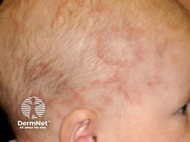
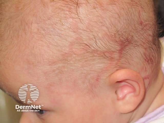
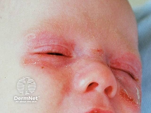
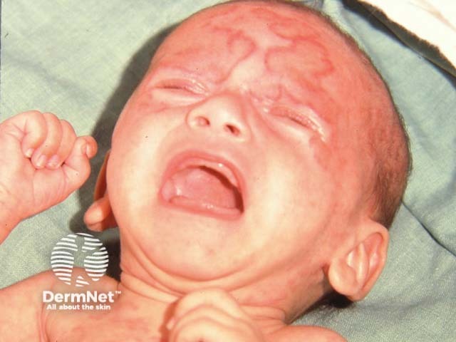
Skin changes may be present at birth or becomes apparent within 3 months of birth, typically soon after first sun exposure.
Neonatal cardiac lupus erythematosus can be complicated by endocardial fibroelastosis and dilated cardiomyopathy with significant morbidity and mortality.
Neonatal lupus erythematosus should be suspected clinically in a baby with congenital heart block and/or typical rash, and confirmed on testing of both mother and baby for ANA, Ro and La (ENA) autoantibodies.
Blood tests should also include a full blood count and liver function tests.
ECG is recommended if heart block has not already been diagnosed before birth on ultrasound as an unusually slow heart rate in the 16–26th week of gestation (second or third trimester).
Skin biopsy is not usually required in the presence of typical clinical features. Histology is reported as showing basal layer vacuolar changes also involving the adnexae, with a superficial and deep periadnexal and perivascular lymphocytic infiltrate.
Hydroxychloroquine is reported to reduce the risk of cardiac neonatal LE in pregnant women with Ro and/or La autoantibodies. Initial studies did not seem to show any benefit for non-cardiac manifestations of neonatal lupus. However, a subsequent large case series reported a reduced risk of cutaneous neonatal lupus with a later onset of the rash when hydroxychloroquine was taken for SLE.
Sun protection reduces the risk of the skin rash. Topical steroid may be applied to the skin lesions.
A permanent pacemaker is required for complete heart block, which is often detectable before birth.
The cutaneous features of neonatal lupus erythematosus slowly resolve over 6–12 months as the maternal antibodies clear from the baby's circulation. Mild epidermal atrophy, telangiectases, and dyspigmentation may persist, particularly if the skin lesions were very inflammatory. Liver, blood, and neurological changes also resolve in a similar timeframe.
Cardiac symptoms do not self-resolve, and the mortality in babies with heart block is up to 20% despite pacemaker implantation.
Affected children should be followed up into early adult life as there is an increased risk of developing rheumatic or other autoimmune diseases.
The risk of neonatal lupus erythematosus in subsequent pregnancies is up to 50%. Subsequent pregnancies should therefore be monitored with serial echocardiograms. Follow-up of asymptomatic mothers is important due to the risk of subsequent connective tissue disease such as SLE.
An AI summary will appear based on your search term using data from all of the topic pages across the entire DermNet site.
Show more