
ADVERTISEMENT
You do not have any notes added to this page yet
Introduction
Demographics
Causes
Clinical features
Complications
Diagnosis
Differential diagnoses
Treatment
Outcome
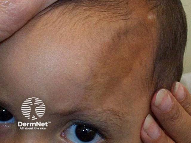
Morphoea en coup de sabre is a variant of linear morphoea (a localised form of scleroderma) restricted to the frontoparietal region (forehead). The name ‘en coup de sabre’ (the blow of a sword) derives from the characteristic scar that indents the skin of the scalp and the underlying bone. Morphoea en coup de sabre also can cut into the brain, causing neurological abnormalities and vision problems.
Morphoea en coup de sabre is also known as frontoparietal linear morphoea (American spelling morphea), linear morphoea en coup de sabre, or just 'en coup de sabre'.
Morphoea en coup de sabre mostly occurs in children (67%), with a slight predominance in girls (about 2:1). It usually first appears in the first two decades of life. The average age of onset was 13.6 years of age in a series of 41 patients reported from the Mayo Clinic over a 20 year period, compared to 32 patients in Wisconsin presenting at 6.9 years of age (range 1-15 years).
The exact cause of morphoea en coup de sabre is unknown. It is believed to be an autoimmune inflammatory disease. There is probably a genetic predisposition, although the specific genetic factors are unknown. The trigger initiating the inflammation is usually unknown, but has been reported to include trauma, radiotherapy, and herpes zoster ophthalmicus. The resulting inflammatory response causes the characteristic damage.
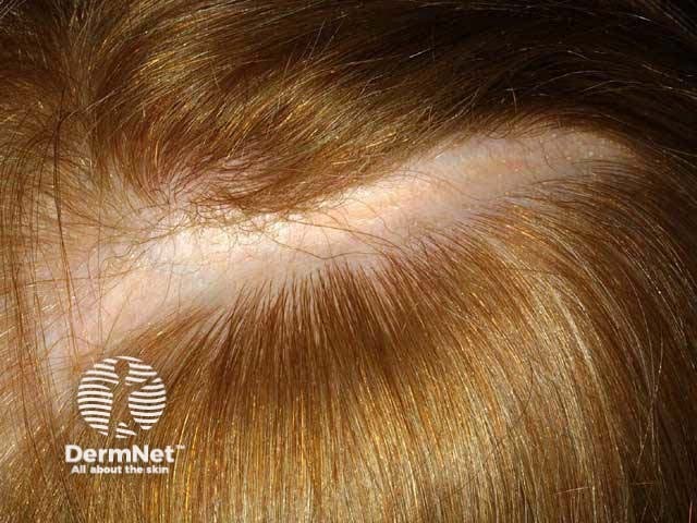
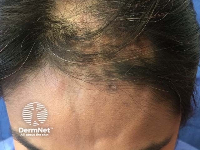
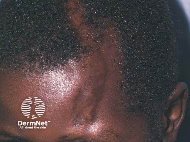
The clinical features of morphoea en coup de sabre evolve over months to years. En coup de sabre begins with a slightly hyperpigmented or hypopigmented streak on the forehead. Other sites may also be involved including the nose, cheek, lip, and neck. Although morphoea en coup de sabre is usually unilateral, changes can affect both sides of the face. Typically the skin becomes ivory in colour with a violaceous margin. The streak becomes indurated, evolving into a scar. The underlying scalp and sometimes even the skull indent which can result in facial asymmetry. As the scar grows, it reaches past the hairline into the scalp, causing linear cicatricial alopecia.
The complications of morphoea en coup de sabre include:
The diagnosis of morphoea en coup de sabre is usually made clinically by noting the characteristic forehead and scalp features. Dermoscopy shows thickened telangiectatic vessels due to inflammation, fibrosis, and loss of follicles.
If necessary, a skin biopsy can distinguish en coup de sabre from similar conditions. The histology shows the classical pathological features of scleroderma.
Negative tests for anti-endothelial antibodies, antinuclear antibodies, and anti-Scl-70 can rule out other connective tissue diseases.
Imaging may include ultrasound, plain X-rays, and computed tomography (CT) scans. Magnetic resonance imaging (MRI) may show skull abnormalities, focal brain atrophy, calcification, and white matter lesions.
Diagnosis can be difficult during the early phase when the morphoea is no more than a pigmented line. Other conditions confused with morphoea en coup de sabre may include:
The goal of treatment is to prevent progression of the en coup de sabre. The most commonly used treatment is a combination of methotrexate with oral corticosteroids. There are no published guidelines for treatment of morphoea en coup de sabre, and no standard of care. A wide range of other treatments have been reported in individual case reports.
Once morphoea en coup de sabre is burnt out, the atrophic skin and underlying structures may undergo surgical repair. Techniques may include:
The natural history of morphoea en coup de sabre is to progress slowly over many years, then to be self-limiting. There are three phases.
The active phase of morphoea en coup de sabre features areas of active sclerosis with erythematous or violaceous borders, with or without expansion of the plaque. The goal of treatment during the active phase is to slow or stop progression with immunosuppressants.
The regression phase of morphoea en coup de sabre retains persistent sclerosis without erythema or a violaceous border. The loss of redness indicates regression.
The burnt-out phase of morphoea en coup de sabre demonstrates no sclerosis and no erythema. The burnt-out phase signals that the course of en coup de sabre is complete, and no further damage will occur.
An AI summary will appear based on your search term using data from all of the topic pages across the entire DermNet site.
Show more