Lymphocytic thrombophilic arteritis is a primary lymphocytic vasculitis. It presents with livedo racemosa or macularhyperpigmentation. It has distinct histological findings affecting small to medium-sized arteries in the deep dermis and superficial subcutis [1].
It is also known as macular lymphocytic arteritis.
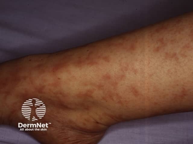
Livedo racemosa
Who gets lymphocytic thrombophilic arteritis?
Lymphocytic thrombophilic arteritis shows a female predominance, with a median age of 39 years (range 6-73) [2].
What causes lymphocytic thrombophilic arteritis?
The exact cause of lymphocytic thrombophilic arteritis is unknown. It has been proposed that the underlying lymphocytic endovasculitis drives a localised thrombophilia, represented by the intraluminal fibrin ring seen on histology, without any evidence of destruction of the vessel wall [1].
Some cases have been associated with autoimmuneantibodies or heterozygosity for prothrombotic mutations, suggesting that autoimmunological and thrombophilic factors may contribute.
Antiphospholipid antibodies have been detected in some cases, but the low titres and lack of systemic features argue against antiphospholipid syndrome contributing significantly to its pathogenesis [1,3].
Low to moderate levels of antinuclear antibodies (ANA) have been reported, but there was no evidence of systemic connective tissue disease in these cases [1,4].
What are the clinical features of lymphocytic thrombophilic arteritis?
The clinical features of lymphocytic thrombophilic arteritis include:
Persistentlivedo racemosa or macular hyperpigmentation typically affecting the lower limbs; the upper limbs can be affected to a lesser extent. This is non-blanching and may form an annular pattern.
How is lymphocytic thrombophilic arteritis diagnosed?
The diagnosis of lymphocytic thrombophilic arteritis is based on a combination of typical clinical features supported by distinct histological findings [1].
A dense inflammatoryinfiltrate is found in the muscular vessel wall, affecting the small and medium-sized arteries of the deep dermis, junction of the dermis and subcutis, or superficial subcutis.
The inflammatory infiltrate consists predominantly of mononuclear cells, mainly lymphocytes and some histiocytes.
Neutrophils and eosinophils are scant or absent.
A concentrichyalinised fibrin ring involves the entire periphery of the lumina of the affected vessels.
Signs or symptoms of systemic vasculitis are also absent (see Cutaneous vasculitis) [2].
What is the differential diagnosis for lymphocytic thrombophilic arteritis?
Lymphocytic thrombophilic arteritis can be confused with several other vascularocclusive conditions showing livedo reticularis.
Cutaneous polyarteritis nodosa
Cutaneous polyarteritis nodosa is characterised by painful subcutaneous nodules and ulceration, features that are uncommon in lymphocytic thrombophilic arteritis [7].
Livedo in cutaneous polyarteritis nodosa is localised around the ulcers with an irregular starburst pattern [7].
Early-stage cutaneous polyarteritis nodosa shows neutrophilicinfiltration involving the muscular layer of medium-sized and small arteries, with more mononuclear cell involvement in the later stages [7].
There is thrombosis without significant inflammation in the vessel wall.
Antiphospholipid syndrome is characterised by recurrent thrombosis, miscarriages, and thrombocytopenia [9].
Sneddon syndrome
The pattern of livedo racemosa in Sneddon syndrome is broader compared to lymphocytic thrombophilic arteritis [3].
The livedo is more proximal in Sneddon syndrome, involving the trunk and back; the extremities are rarely involved.
The diagnosis of Sneddon syndrome requires cerebrovascular involvement [10].
What is the treatment for lymphocytic thrombophilic arteritis?
Lymphocytic thrombophilic arteritis tends to show an indolent course with persistent asymptomaticlivedo racemosa or macular hyperpigmentation [1]. As most lesions are asymptomatic, treatment is not always required [2].
Treatment options that have shown a beneficial response include:
Given the rarity of lymphocytic thrombophilic arteritis and inconsistent treatment responses, there is no current consensus on treatment recommendations.
What is the outcome for lymphocytic thrombophilic arteritis?
Lymphocytic thrombophilic arteritis tends to follow a chronic indolent course with no progression to systemic vasculitis [1,2]. Ongoing follow-up is advisable as there have been rare reports of later systemic vasculitis [11]. The long-term prognosis remains unclear.
References
Lee JS, Kossard S, McGrath MA. Lymphocytic thrombophilic arteritis: a newly described medium-sized vessel arteritis of the skin. Arch Dermatol 2008; 144: 1175–82. DOI: 10.1001/archderm.144.9.1175. PubMed
Vakili S, Zampella JG, Kwatra SG, Blanck J, Loss M. Lymphocytic thrombophilic arteritis: a review. J Clin Rheumatol 2019; 25: 147–52. DOI: 10.1097/RHU.0000000000000846. PubMed
Gupta S, Mar A, Dowling JP, Cowen P. Lymphocytic thrombophilic arteritis presenting as localized livedo racemosa. Australas J Dermatol 2011; 52: 52–5. DOI: 10.1111/j.1440-0960.2010.00673.x. PubMed
Sadahira C, Yoshida T, Matsuoka Y, Takai I, Noda M, Kubota Y. Macular arteritis in Japanese patients. J Am Acad Dermatol 2005; 52: 364–6. DOI: 10.1016/j.jaad.2004.08.015. PubMed
Kelly RI, Wee E, Tancharoen C, Tam MM, Balta S, Williams RA. Three cases of lymphocytic thrombophilic arteritis presenting with an annular eruption. Australas J Dermatol 2018; 59: e127–32. DOI: 10.1111/ajd.12679. PubMed
Llamas-Velasco M, García-Martín P, Sánchez-Pérez J, Sotomayor E, Fraga J, García-Diez A. Macular lymphocytic arteritis: first clinical presentation with ulcers. J Cutan Pathol 2013; 40: 424–7. DOI: 10.1111/cup.12094. PubMed
Morgan AJ, Schwartz RA. Cutaneous polyarteritis nodosa: a comprehensive review. Int J Dermatol 2010; 49: 750–6. DOI: 10.1111/j.1365-4632.2010.04522.x. PubMed
Alavi A, Hafner J, Dutz JP, et al. Livedoid vasculopathy: an in-depth analysis using a modified Delphi approach. J Am Acad Dermatol 2013; 69: 1033–42.e1. DOI: 10.1016/j.jaad.2013.07.019. PubMed
Miyakis S, Lockshin MD, Atsumi T, et al. International consensus statement on an update of the classification criteria for definite antiphospholipid syndrome (APS). J Thromb Haemost 2006; 4: 295–306. DOI: 10.1111/j.1538-7836.2006.01753.x. PubMed
Wee E, Nikpour M, Balta S, Williams RA, Kelly RI. Lymphocytic thrombophilic arteritis complicated by systemic involvement. Australas J Dermatol 2018; 59: 223–5. DOI: 10.1111/ajd.12798. PubMed