
ADVERTISEMENT
You do not have any notes added to this page yet
Introduction Demographics Causes Clinical features Complications Diagnosis Differential diagnoses Treatment Outcome
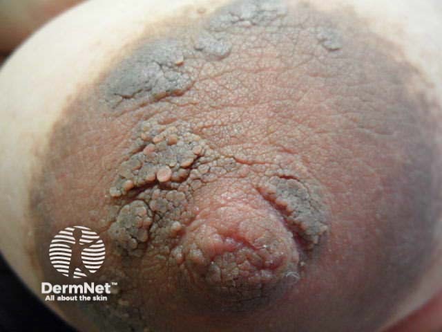
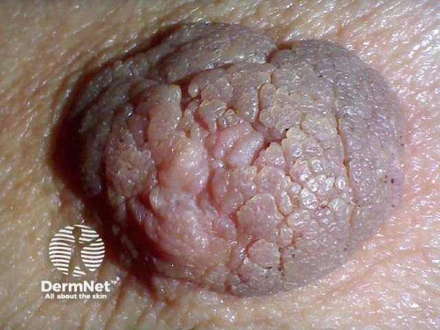
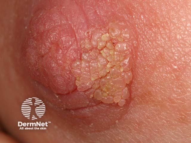
Hyperkeratosis of the nipple and areola is a warty pigmented thickening of the nipples and areolae [1–4]. It can be primary (idiopathic) or secondary to another disorder (see differential diagnosis).
Hyperkeratosis of the nipple and areola is also called naevoid hyperkeratosis and hyperkeratosis areolae mammae naeviformis.
Primary hyperkeratosis of the nipple typically presents in adolescent females, with the rate in males being much lower [1,2,3]. No ethnic or geographical links have been reported.
The exact cause of hyperkeratosis of the nipple is not known. Although no hormonal alterations have been found, an endocrinological cause has been proposed due to its association with the female sex, oestrogen therapy, and pregnancy [1].
The clinical features associated with hyperkeratosis of the nipple follow.
Hyperkeratosis of the nipple may cause embarrassment and cosmetic concerns [4]. There are reported cases of difficulty breast-feeding from the affected breast [1].
Diagnosis of hyperkeratosis of the nipple relies upon the clinical presentation of the hyperkeratotic plaque(s) and findings on skin biopsy if the presentation is atypical.
Histopathological characteristics of hyperkeratosis of the nipple are:
Secondary hyperkeratosis of the nipple [1,2,4] is usually unilateral. Examples include:
Unilateral hyperkeratosis of the nipple associated with pain, bleeding, ulceration, discharge, or nipple retraction should be investigated for breast cancer with breast examination, mammogram, and biopsy.
Bilateral secondary hyperkeratosis of the nipple and areola may occur with:
Untreated hyperkeratosis of the nipple does not usually interfere with the normal breast function. However, treatment of the lesion is sometimes requested for cosmetic reasons [3,4].
Cryotherapy is a suitable first-line treatment for hyperkeratosis of the nipple often with a cosmetically satisfactory result [3,4]. It may need to be repeated. Hypopigmentation is the main long-term risk of cryotherapy.
Medical treatments do not remove the lesions permanently but keratolytic agents (such as salicylic acid, lactic acid, or urea), topical retinoids, and calcipotriol, may be helpful [2,4].
Skin surgery or laser resurfacing is sometimes undertaken but may be cosmetically unsatisfactory [4].
Without treatment, hyperkeratosis of the nipple persists.
Cryotherapy or surgical excision reduces recurrence [3,4].
An AI summary will appear based on your search term using data from all of the topic pages across the entire DermNet site.
Show more