Author: Rajan Ramji, 5th Year Medical Student, University of Auckland, New Zealand; Chief Editor: Hon A/Prof Amanda Oakley, Dermatologist, Hamilton, New Zealand. April 2016.
A leiomyoma is a benigntumour composed of smooth muscle. It is capable of arising wherever smooth muscle is present. One form of leiomyoma arises from uterine smooth muscle, and is otherwise known as uterine fibroids.
Cutaneous leiomyomas may be classified into three types:
Piloleiomyoma
Angioleiomyoma
Genital leiomyoma
Each type arises from smooth muscle in specific tissues or organs and has distinct clinical or histological features.
Piloleiomyoma
Originates from the arrector pili muscle of the pilosebaceous unit
Solitary or multiple lesions
Multiple lesions may occur sporadically or are inherited alongside uterine fibroids in an autosomal dominant pattern as part of hereditary leiomyomatosis and renal cell cancer syndrome or as part of Reed syndrome (multiple cutaneous and uterine leiomyomatosis)
Angioleiomyoma
Originates from vascular wall smooth muscle tissue
Typically occurs as a solitary lesion
Genital leiomyoma
Originates from the dartos muscle in the scrotum or labium major, or from erectile muscle in nipples
Typically occurs as a solitary lesion
Least common of the cutaneous leiomyoma
Who gets leiomyomas?
Uterine leiomyomas represent 95% of all reported leiomyomas. Cutaneous leiomyomas represent 75% of all extra-uterine leiomyomas.
Incidence is largely unrelated to race.
The majority of solitary cutaneous leiomyomas occur in adulthood. Multiple piloleiomyomas typically occur between the ages of 10 and 30 years.
Excluding autosomal dominant syndromes, piloleiomyoma incidence is equal in men and women. Angioleiomyomas are generally more common in women than men (2:1) although cavernous and venous subtypes are more common in men.
What are the clinical features of leiomyomas?
Leiomyomas are often painful.
Pain may be spontaneous or triggered by physical and/or emotional stimuli, including cold temperature or pressure. Menses or pregnancy may also act as triggers
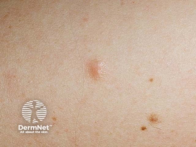
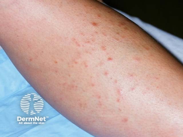
Piloleiomyomas
Likely to present with associated pain
Typically tender, mobile hyperpigmented or reddish brown nodules (≤ 2cm diameter) with a smooth surface and firm consistency
Solitary lesions typically in the lower extremities while multiple piloleiomyomas appear anywhere in a variety of distribution patterns
Angioleiomyomas
60% reported to be painful
Tenderness is less common than in piloleiomyomas
Well circumscribed, skin coloured, solitary nodules ≤ 4cm in diameter usually found on the lower legs. Uncommonly on head, trunk, hands or mouth.
Genital leiomyomas
Typically non-tender and painless
Firm, solitary, skin coloured, mobile nodule on the vulva, scrotum or nipple. Size is more variable than other types; vulval and scrotal lesions are typically larger.
Leiomyomas
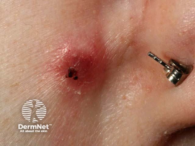
How are cutaneous leiomyomas usually diagnosed?
Cutaneous leiomyomas are usually diagnosed by skin biopsy. Each type of leiomyoma has unique histology.
How are leiomyomas treated?
Surgical excision is the definitive treatment for single lesions. Multiple cutaneous leiomyomas have a high rate of recurrence (~50%) within weeks to years – especially if part of HLRCC or Reed syndrome.
Medical treatment is not curative, but nifedipine, phenoxybenzamine and gabapentin may provide relief of pain.
References
Alam M, Rabinowitz AD, Engler DE. Gabapentin treatment of multiple piloleiomyoma-related pain. J Am Acad Dermatol. 2002 Feb. 46(2 Suppl Case Reports):S27–9. PubMed
Batchelor RJ, Lyon CC, Highet AS. Successful treatment of pain in two patients with cutaneous leiomyomata with the oral alpha-1 adrenoceptor antagonist, doxazosin. Br J Dermatol. 2004 Apr. 150(4):775–6. PubMed
Brooks JK, Nikitakis NG, Goodman NJ, Levy BA. Clinicopathologic characterization of oral angioleiomyomas. Oral Surg Oral Med Oral Pathol Oral Radiol Endod. 2002 Aug. 94(2):221–7. PubMed
Gokdemir G, Sakiz D, Koslu A. Multiple cutaneous leiomyomas of the nipple.J Eur Acad Dermatol Venereol. 2006 Apr. 20(4):468–9. PubMed
Holst VA, Junkins-Hopkins JM, Elenitsas R. Cutaneous smooth muscle neoplasms: clinical features, histologic findings, and treatment options. J Am Acad Dermatol. 2002 Apr. 46(4):477–90; quiz, 491–4. PubMed
Khachemoune A, Rodriguez C, Lyle S, Jiang SB. Genital leiomyoma: surgical excision for both diagnosis and treatment of a unilateral leiomyoma of the male nipple. Dermatol Online J. 2005 Jan 1;11(1). PubMed
Kudur MH. A generalized multiple cutaneous piloleiomyomatosis in a young male: Rare case report. Indian J Dermatol. 2013 May;58(3):245. PubMed
Malhotra P, Walia H, Singh A, Ramesh V. Leiomyoma cutis: a clinicopathological series of 37 cases. Indian J Dermatol. 2010 Oct 1;55(4):337. PubMed
Nagata S, Nishimura H, Uchida M, Hayabuchi N, Zenmyou M, Fukahori S. Giant angioleiomyoma in extremity: report of two cases. Magn Reson Med Sci. 2006 Jul. 5(2):113–8. PubMed
Ramesh P, Annapureddy SR, Khan F, Sutaria PD. Angioleiomyoma: a clinical, pathological and radiological review. Int J Clin Pract. 2004 Jun. 58(6):587–91. PubMed
Scheinfeld N. The role of gabapentin in treating diseases with cutaneous manifestations and pain. Int J Dermatol. 2003 Jun. 42(6):491–5. PubMed
Yagi K, Hamada Y, Yasui N. A leiomyoma arising from the deep palmar arterial arch. J Hand Surg [Br]. Dec 2006. 31(6):680–2. PubMed