Authors: Dr Charlotte Foster, Anatomical Pathology Registrar, Tauranga, New Zealand; Dr Ben Tallon, Consultant Dermatopathologist, Tauranga, New Zealand. Copy edited by Gus Mitchell. September 2021.
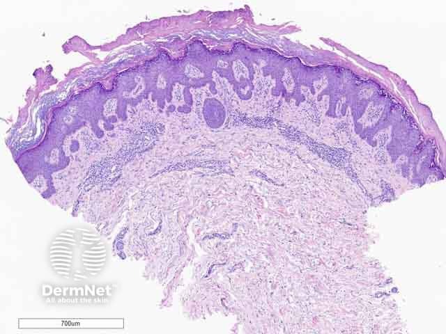
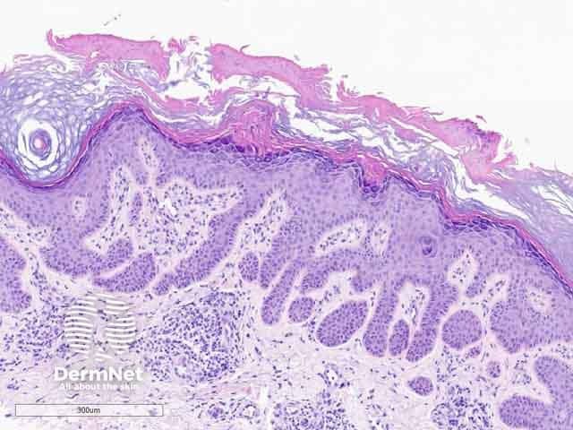
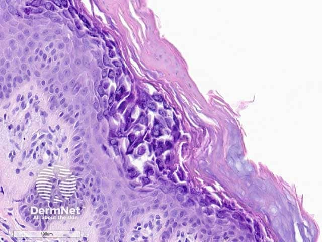
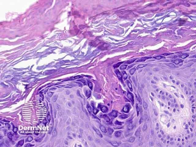
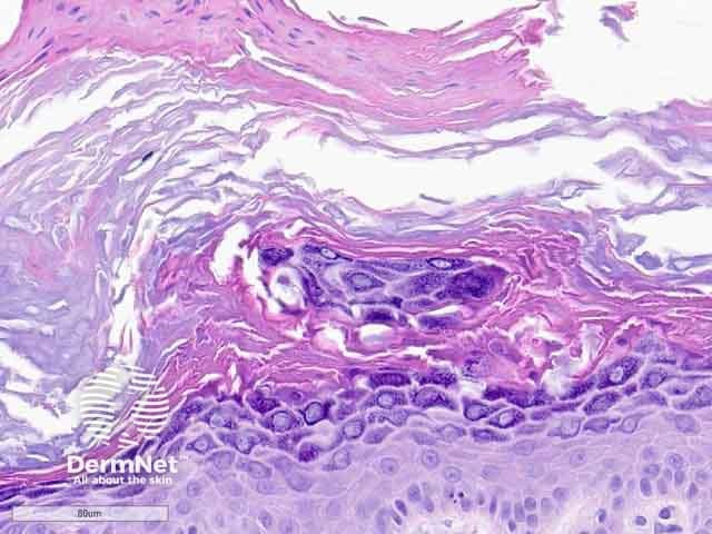
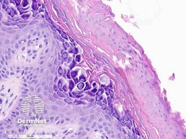
Hypergranulotic dyscornification is reminiscent of verruca vulgaris. The lesion may be exoendophytic (figure 1). There are finger-like projections of epidermalhyperplasia and hypergranulosis with clustered keratohyalin granules. The entire lesion shows compact orthokeratosis underneath a laminated and basket-weave stratum corneum (figure 2). The key feature is the corneocytes in the stratum corneum which appear rounded, glassy, and eosinophilic (figures 3–6). Parakeratosis is also usually present. There is a variable underlying inflammatorylymphocyticinfiltrate in the upper dermis.
Figure 1: low power
Figure 2: medium power
Figure 3: high power
Figure 4: high power
Figure 5: high power
Figure 6: high power
Differential diagnosis of hypergranulotic dyscornification pathology
Epidermolytic hyperkeratosis. The keratohyalin granules are not as thick and clumped, but instead small and dotted in epidermolytichyperkeratosis. The eosinophilic corneocytes seen in hypergranulocytic dyscornfication are also not present. There is usually reticular degeneration present in epidermolytic hyperkeratosis.
Verruca vulgaris. Koilocytes are present in verruca vulgaris and not seen in hypergranulotic dyscornification.
Seborrhoeic keratosis. Hornpseudocysts will usually be present in seborrhoeickeratosis and absent in hypergranulotic dyscornification.
Bibliography
Gesheva A, Pitch M, Rosamilia L, Hossler E. Keratotic papule on the abdomen. Cutis. 2020;105(5):222–31. Journal
Reichel M. Hypergranulotic dyscornification: a distinctive histologic pattern of maturation of epidermal epithelium present in solitary keratoses. Am J Dermatopathol. 1999;21(1):21–4. doi:10.1097/00000372-199902000-00004 PubMed
Roy SF, Ko CJ, Moeckel GW, Mcniff JM. Hypergranulotic dyscornification: 30 cases of a striking epithelial reaction pattern. J Cutan Pathol. 2019;46(10):742–7. doi:10.1111/cup.13522. PubMed