Verruca vulgaris (common viral wart) is a keratoticlesion caused by specific human papillomavirus (HPV) types.
Histology of verruca vulgaris
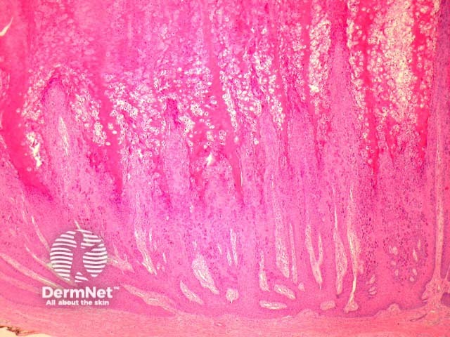
Histopathologic examination of verruca vulgaris reveals a markedly papillomatousepidermis with hypergranulomatosis and overlying tiers of parakeratosis (figure 1).
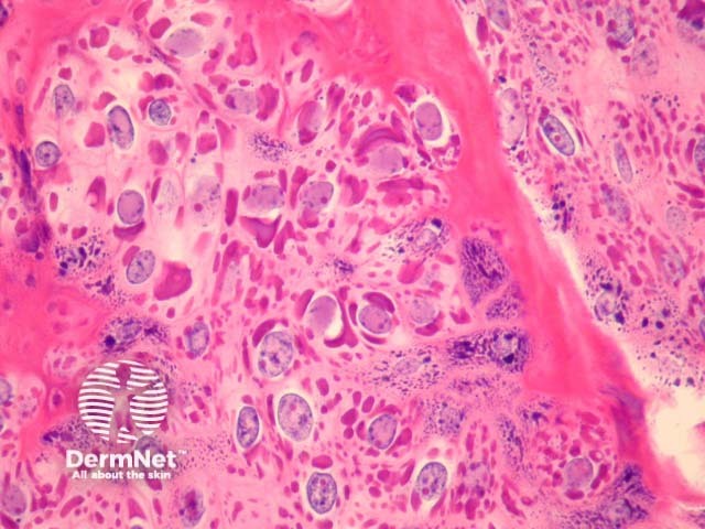
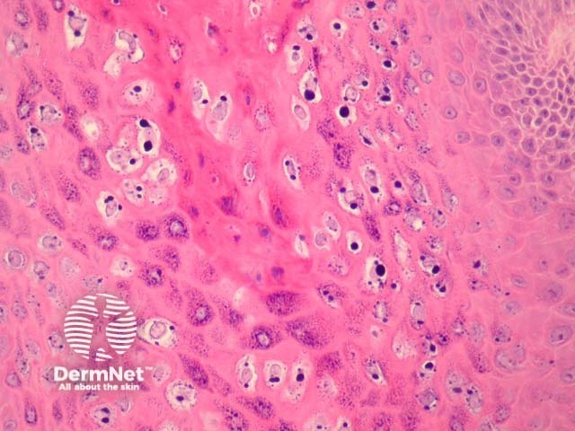
The upper epidermis may contain large pink inclusions (figure 2), particularly in cases arising on acral skin. Other lesions show smaller basophilicgranules (figure 3). Characteristic vacuolatedkeratinocytes (koilocytes), which have a small shrunken nucleus surrounded by a perinuclear halos, are seen in the upper epidermis (figure 3).
Figure 1
Figure 2
Figure 3
Special stains for verruca vulgaris
Special stains are not required to make the diagnosis. PCR may be used to identify the HPV type.
Differential diagnosis of verruca vulgaris pathology
Seborrhoeic keratosis – Distinction can be difficult for older verrucas in which the viropathic effect is difficult to appreciate. Some pathologists use the term “verrucal keratosis” or similar in cases where a clear distinction cannot be made.
Molluscum contagiosum – The large viral inclusions of molluscum should not be confused with the very large basophilic inclusions which may be seen in some verrucae (figure 2). Large HPV inclusions are usually seen in lesions occurring on acral skin, an exceedingly uncommon site for molluscum contagiosum.
References
Weedon’s Skin Pathology (Third edition, 2010). David Weedon
Pathology of the Skin (Fourth edition, 2012). McKee PH, J. Calonje JE, Granter SR