
ADVERTISEMENT
You do not have any notes added to this page yet
Introduction
Demographics
Diagnosis
Differential diagnoses
Treatment
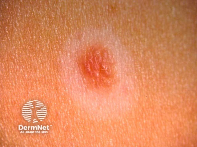
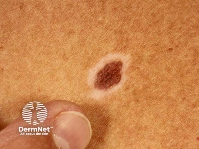
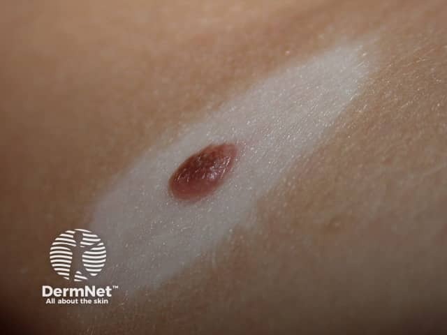
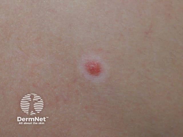
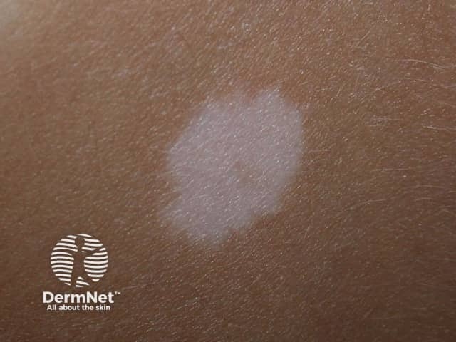
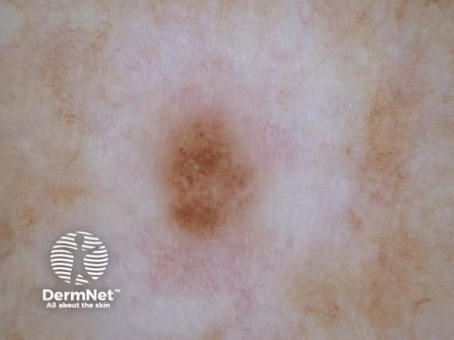
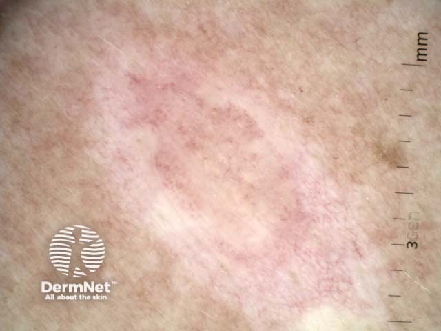
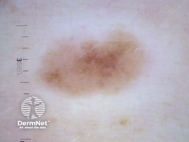
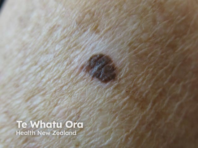
A halo naevus (US spelling, nevus) is an otherwise normal mole with a white ring, or halo, around it. The central dark brown naevus fades from dark brown to light brown to pink, eventually disappearing completely.
Halo naevus is also known as:
Halo naevi (US plural, nevi) are not uncommon, with an estimated prevalence of 1% of the white-skinned population. They are usually seen in healthy children or young adults of either sex. They can occur at an older age too.
Halos can be seen as part of a more generalised pigment loss, vitiligo, and halo naevi may also be associated with another autoimmune disease.
They may rarely arise as a reaction to advanced melanoma and in patients with metastatic melanoma treated with targeted therapy or immune checkpoint inhibitors such as pembrolizumab or nivolumab.
Why halo naevi develop is not fully understood. They are currently classified as autoimmune in origin.
The onset of a halo naevus may be triggered by sunburn or local trauma, which causes the mole to be recognised by the immune system as foreign, resulting in an attack by circulating antibodies and CD8+ T lymphocytes. The reaction also affects the normal skin around the mole, which also has pigment cells in it, causing depigmentation.
Development of halo naevi has also been associated with psychosocial stress.
A solitary halo naevus or multiple halo naevi are most often found on the trunk. They are less common on the head and are rare on the limbs. The affected naevi are dermal naevi that are congenital or have arisen during childhood. Halo naevi may follow the Koebner phenomenon, arising within a mole that has been injured in some way.
The white halo is usually about 0.5–1.0 cm wide and is symmetrical (round or oval in shape). The halos develop at intervals round one or several moles, but not around all of them.
There are four stages of a halo naevus. It may take several years to complete the cycle. Multiple halo naevi can be at different stages.
See more images of halo naevi.
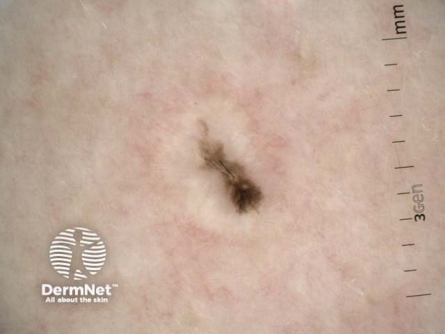
Halo naevus is a clinical diagnosis. Dermoscopy may be used to evaluate the structure and colour of the naevus.
A full skin examination should be performed (especially in adults), as rarely, halo naevi can be triggered by the presence of a melanoma elsewhere.
Occasionally, if it has atypical features such as irregularity in structure or colour, excision of a solitary halo naevus is recommended to make sure it is benign. Histology reveals a band-like lymphohistiocytic infiltrate in the dermis under the naevus.
The differential diagnosis of halo naevus includes:
Where there is doubt about the diagnosis, the whole naevus should be excised for histopathological examination. Partial biopsy could be misleading.

Apart from an explanation, no treatment is normally required for a typical halo naevus.
The white skin of a halo naevus will burn particularly easily in the sun because it is missing protective melanin pigment. Cover up or apply sunscreen during summer to prevent sunburn.
Surgery is not usually necessary but may be recommended if there are atypical features such as irregularity in the structure of the naevus.
An AI summary will appear based on your search term using data from all of the topic pages across the entire DermNet site.
Show more