A fistula is an abnormal channel leading between two cavities or surfaces which may drain a fluid material such as saliva or pus. An example would be from the mouth (oral cavity) to the skin surface, usually of the face or neck, and this specific type is called an orocutaneous fistula.
A sinus has one open draining end and the channel ends in a blind ending. An example would be a dental sinus draining from a dental abscess to either the inside of the mouth or the skin.
The words however are often used interchangeably.
Fistulas and sinuses of the neck and face: classification
Fistulas and sinuses of the neck and face may be classified by cause.
Developmental
Fistulas and sinuses due to developmental causes are usually present at birth.
Thyroglossal duct cyst – the most common developmental cyst in the neck. The cyst characteristically moves upwards when the tongue is poked out or with swallowing. It may burst to form a sinus which usually opens just below the hyoid bone in the midline of the neck. It drains mucus. Treatment is surgical (Sistrunk procedure) but 10% recur.
Branchial cleft cyst (lateral branchial arch cyst) – the most common developmental cyst of the side of the neck. A sinus may drain mucus or pus following rupture of an abscess. It usually opens on the side of the neck just above the junction of the collarbone and breast bone (sternoclavicular joint), in front of the sternocleidomastoid muscle. There may also be an associated sinus draining into the pharynx.
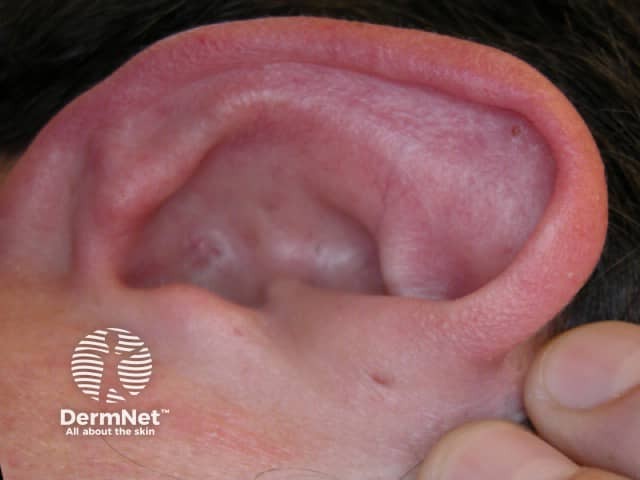
Preauricular pits and sinuses – these are common, affecting 1% of the population, particularly Asians and blacks. 25% are bilateral in front of both ears. The sinus opening (pit) is usually located just in front of the upper part of the ear where the cartilage of the ear rim (helix) meets the facial skin. They are asymptomatic unless infected (uncommon), when they become red, sore and may discharge pus.
Preauricular sinus
Medial nasal dermal fistulae
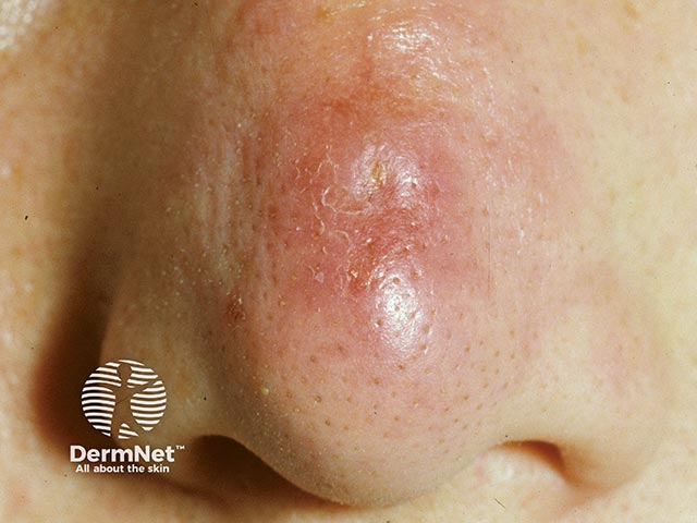
These rare lesions are distinctive - they present with a dimple in the midline of the dorsal nose at the junction of the bony and cartilaginous portions of the nose. Hairs may emanate from them. It is a congenital anomaly formed from the sequestration of fetal ectodermal cells in the process of nasal fusion. The lesion may be a small dimple, or a sinus that can extend down towards the skull and even the dura. Discharge and recurrent infective episodes can indicate their presence. Detailed radiological assessment is needed prior to considering excision of the sinus tract
Recurrently inflamed area at the junction of the cartilaginous and bony nose. It marks the outlet of a median nasal dermal fistula. (ND-patient1)
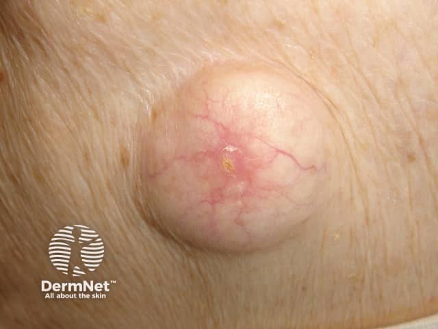
Cysts
Cysts are lumps in the skin containing fluctuant contents. They may have an opening to the skin surface.
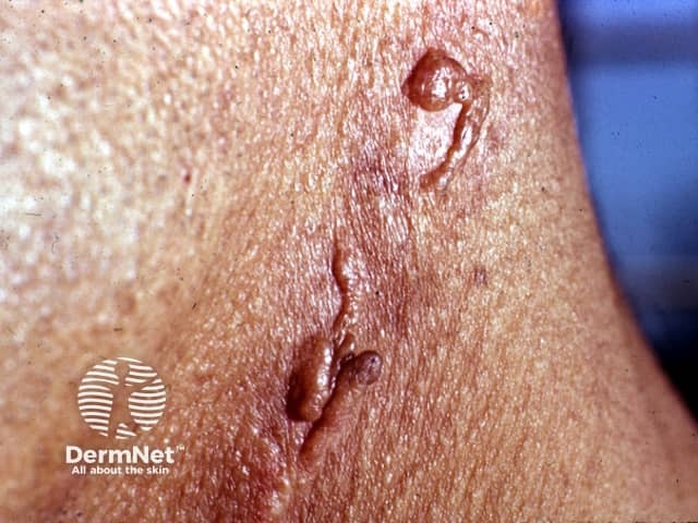
Bone infection Chronicosteomyelitis – most commonly associated with poorly controlled diabetes mellitus or following radiotherapy to the jaw for cancer or Paget disease of the bone. It may also complicate a chronic dental infection.
In addition to careful history and examination, one or more of the following tests will usually be required to confirm the diagnosis and determine the cause:
passing a probe into the channel
radiology – may include plain x-rays, x-rays using contrast medium, CT or MRI scans
microbiological assessment of swabs or biopsy material