
ADVERTISEMENT
You do not have any notes added to this page yet
Introduction Demographics Causes Clinical features Variation in skin types Complications Diagnosis Differential diagnoses Treatment Prevention Outcome
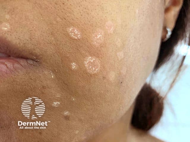
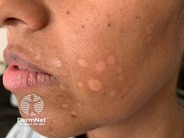
Facial discoid dermatosis (FDD) is a rare dermatosis that presents as discrete and persistent pink-orange papulosquamous lesions isolated to the face.
FDD was first described in 2010 by Ko et al, and since then multiple case reports and series have been described in the literature. It is rare and therefore often misdiagnosed and under-recognised as a clinical entity. It can be notoriously difficult to treat, and often does not respond to conventional treatments, including topical steroids.
It is estimated from case series that FDD affects women more commonly than men, with some authors reporting a male-to-female ratio of 1:5. Similarly, based on limited patient numbers in the literature, it is thought to mainly affect patients from an Asian background (Chinese, Indian, and Turkish cases have been reported).
The aetiology of FDD is currently unknown.
FDD usually presents as discrete, annular, well-demarcated pink-orange plaques and papules that may have a fine superficial scale. It is limited in location to the face and neck, and can present over the course of months to years.
Given limited reported cases, the clinical features across differing types of skin are unknown.
There are no reported complications of FDD, and it is not known to be associated with any underlying medical disease.
Biopsy findings can be non-specific, and may include components such as hyper/parakeratosis, follicular plugging, acanthosis, and a lymphocytic infiltrate in the superficial dermis.
However, a biopsy can be important to rule out other causes such as fungal infections and atypical psoriasis.
The differential diagnosis is broad and can include:
Given the paucity of cases reported in the literature, general treatment measures are unknown.
Numerous treatments have been used across case reports, with varying degrees of success.
Topical treatments that are most commonly tried include topical steroids, vitamin D analogues, and calcineurin inhibitors.
The only oral treatment that has been successfully trialled is low dose acitretin, however this was in combination with topical calcipotriol/betamethasone. This was confirmed in another case report with successful use of topical calcipotriol, after failed courses of: topical miconazole, clobetasone butyrate, tacrolimus; and oral valaciclovir, fluconazole, and clarithromycin.
More recently there has been a case report of successful treatment of FDD with interleukin (IL) 12 and IL-23 inhibitor ustekinumab.
Given the paucity of case reports and understanding of the condition, this is not known.
Prognosis has varied between case reports, but it is known to be difficult to treat and often unresponsive to topical and oral treatments. Successful use of topical calcipotriol, low-dose oral acitretin, and subcutaneous ustekinumab have all been reported in the literature.
An AI summary will appear based on your search term using data from all of the topic pages across the entire DermNet site.
Show more