Blastomycosis-like pyoderma is a form of pyoderma which elicits an impressive verrucousepidermal reaction and draining sinus. Various bacteria have been implicated as possible causative organisms.
Histology of blastomycosis-like pyoderma
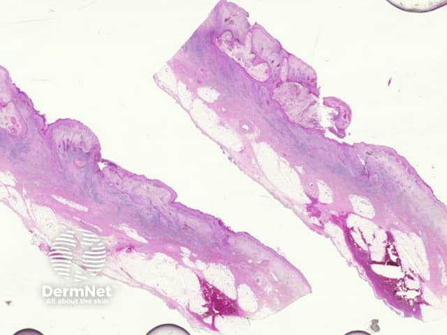
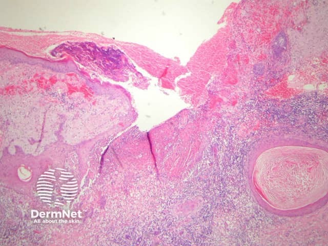
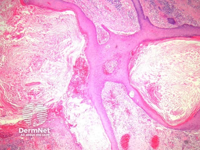
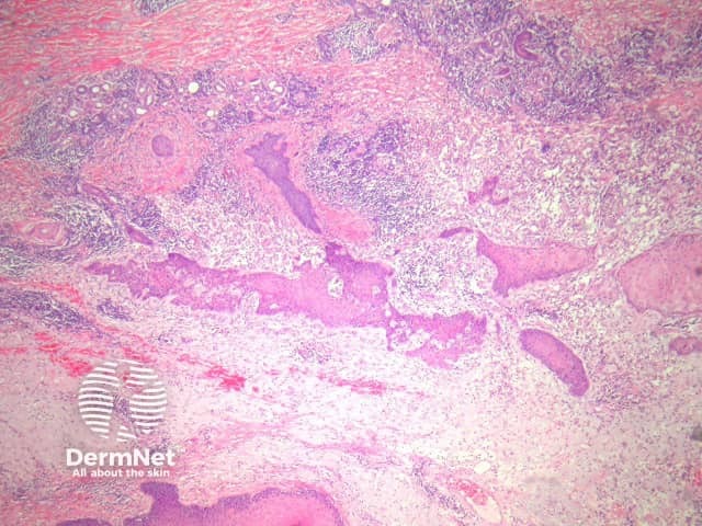
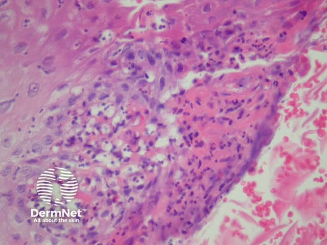
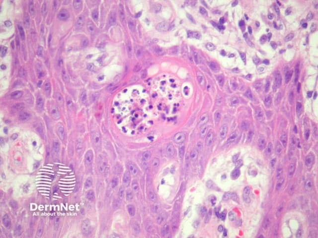
In blastomycosis-like pyoderma, sections usually show a broad verrucous lesion with epidermal papillomatosis which may resemble a keratoacanthoma (figure 1). There may be surface erosion and suppuration (figure 2). Numerous cystic spaces connect with the surface via draining sinuses (figure 3). There may be a pseudo-infiltrative growth pattern as the regenerative epithelium mimics an invasivesquamous cell carcinoma (figure 4). Neutrophilic collections are present in the dermis and within squamous islands (figures 5, 6).
Figure 1
Figure 2
Figure 3
Figure 4
Figure 5
Figure 6
Special studies for blastomycosis-like pyoderma
Gram stain and culture studies may reveal bacteria. Staphylococcus aureus is the most frequent causative agent.
Differential diagnosis of blastomycosis-like pyoderma pathology
Keratoacanthoma – The surface epithelialhyperplasia may be difficult to distinguish from a keratoacanthoma.
Blastomycosis – The reaction pattern is very similar. Special stains for fungal forms should be performed.
Atypical mycobacterial infection/sporotrichosis — these infections may also cause a massive epidermal response. Searching for the causative organisms and correlation with the cultures results can be helpful.
Actinic comedonal plaque — may represent a later stage of blastomycosis-like pyoderma. The inflammation is typically less striking and there is usually impressive solar elastosis.
References
Weedon’s Skin Pathology (Third edition, 2010). David Weedon