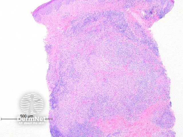
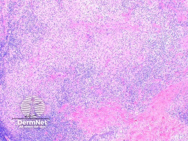
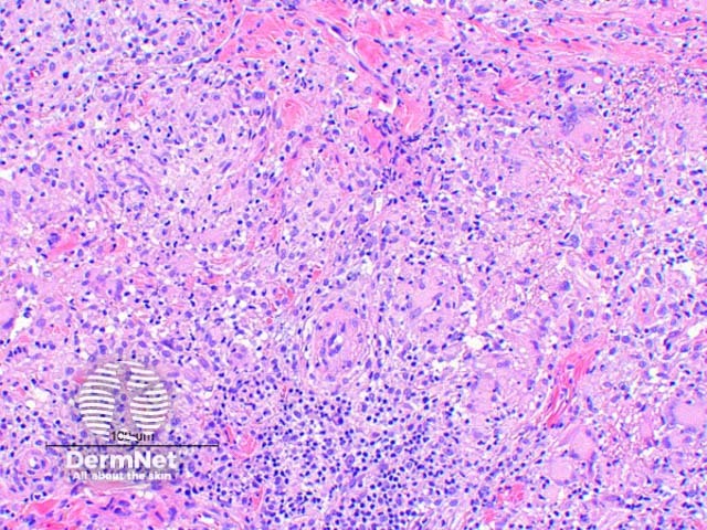
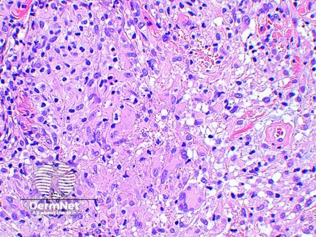
The histologic findings of Mycobacterium marinuminfection vary by the age of the lesion. Scanning power view of well developed lesions demonstrate a granulomatousdermatitis (Figure 1), forming an extensive inflammatorynodularinfiltrate within the dermis. Early lesions may show an acutesuppurative inflammatory process with little granuloma formation. The epidermis may show prominent pseudoepitheliomatoushyperplasia with or without ulceration. There are tuberculoid granulomas with varying degrees of abscess formation (Figure 2). The infiltrate is mixed lymphohistiocytic with multinucleatedgiant cells and scattered neutrophils (Figures 3 and 4).
Figure 1
Figure 2
Figure 3
Figure 4
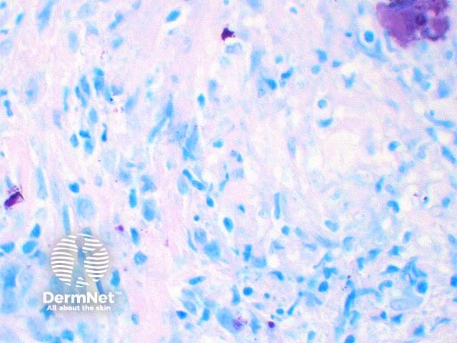
Special stains in Mycobacterium marinum skin infection
Acid fast stains such as Ziehl-Neelsen (Figure 5) reveal bacteria in the majority of cases, but can be negative. Culture at 25-30 Celsius is therefore essential to making a diagnosis in cases with suspicious histology.
Figure 5
Differential diagnosis of Mycobacterium marinum skin infection
Atypicalinfections: Other mycobacterial organisms need to be considered based on histologic findings alone. Neutrophilinfiltrates, interstitial granulomas, small vessel proliferation and high numbers of bacilli are more frequently seen in non-tuberculous (over tuberculous) mycobacterial infections. Cultures studies and PCR are essential for species identification. Deep fungal infections also show suppurative granuloma formation, with fungal elements visible on PAS or silver staining.
Interstitial granuloma annulare: Rare cases have been described where the histology showed an interstitial histiocytic infiltrate resembling interstitial granuloma annulare. Acid fast staining revealed bacterial organisms.
References
Skin Pathology (3rd edition, 2002). Weedon D
Pathology of the Skin (3rd edition, 2005). McKee PH, J. Calonje JE, Granter SR
Travis WD, Travis LB, Roberts GD, Su DW, Weiland LW. The histopathologic spectrum in Mycobacterium marinum infection. Arch Pathol Lab Med. 1985 Dec;109(12):1109–13. PubMed
Min KW, Ko JY, Park CK. Histopathological spectrum of cutaneous tuberculosis and non-tuberculous mycobacterial infections. J Cut Pathol. 2012; 93: 582–95. PubMed
Barr KL, Lowe L, Su LD. Mycobacterium marinum infection simulating interstitial granuloma annulare: a report of two cases. Am J Dermatopathol. 2003 Apr;25(2):148–51. PubMed