Author: Dr Moushumi Das, Medical Registrar, Waikato Hospital, Hamilton, New Zealand. Editor-in-Chief: A/Prof Amanda Oakley, Dermatologist, Hamilton, New Zealand. Copy edited by Gus Mitchell. February 2016. DermNet Revision April 2021
Benign lymphoplasmacytic plaque is a pseudolymphoma in children characterised by one or more chronic skin lesions, either solitary or in linear clusters, that have a characteristic polyclonal plasma cell infiltrate on histology. It was first described in 2009, with few reported cases so far.
Benign lymphoplasmacytic plaque has also been called cutaneous lymphoplasmacytic lymphoma and pretibial lymphoplasmacytic plaque.
Who gets benign lymphoplasmacytic plaque?
Benign lymphoplasmacytic plaque has mostly been reported to affect female children between the ages of 7 and 15 years. Isolated cases have been described in adult males over the age of 60. It seems to predominantly affect people of Caucasian descent, although cases have also been described in Japanese, African, and South Asian (Indian) children.
What causes benign lymphoplasmacytic plaque?
The cause of benign lymphoplasmacytic plaque is unknown. Preceding trauma to the affected area has been noted in a few cases, but no direct correlation has been confirmed.
What are the clinical features of benign lymphoplasmacytic plaque?
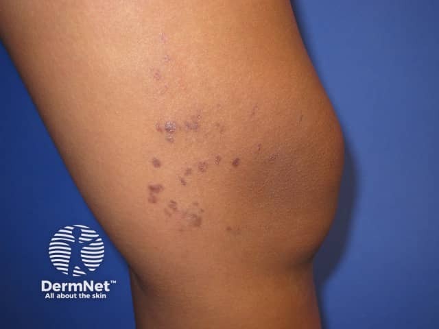
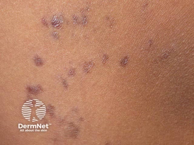
Benign lymphoplasmacytic plaque presents as a solitary chronic, persistent, and asymptomatic skin lesion that is resistant to topical and systemic treatment. Clusters of similar lesions often in a linear pattern may occur.
The mean duration is 3 years.
The lesion is a variable, reddish-brown, painless papule, or a scaly plaque.
They most often affect the lower legs, but may also arise on the trunk, upper limbs, and buttocks.
There are usually no other lesions elsewhere, and there are no systemic symptoms.
Lymphoplasmacytic plaque
Lymphoplasmacytic plaque
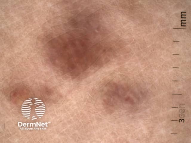
Lymphoplasmacytic plaque: dermatoscopy
How is benign lymphoplasmacytic plaque diagnosed?
A skin biopsy is required as the diagnosis of benign lymphoplasmacytic plaque is made by dermatopathology and clinicopathologic correlation. The defining, compulsory histological findings are:
Dense lymphoplasmacytic infiltrate
The presence of large numbers of plasma cells
The absence of clonality of plasma cells ie, polyclonal plasma cell infiltrate
What is the outcome for benign lymphoplasmacytic plaque?
It is uncertain how benign lymphoplasmacytic plaque progresses long term. Two cases persisting for ten years have been reported with no clinical changes. Spontaneous regression have not been reported.
References
Gilliam AC, Mullen RH, Oviedo G, et al. Isolated benign primary cutaneous plasmacytosis in children: two illustrative cases. Arch Dermatol. 2009;145(3):299-302. doi:10.1001/archdermatol.2008.596.Journal
Harkemanne E, Dargent JL, Roquet-Gravy PP, Bulinckx A. Lymphoplasmacytic plaque in children: case report and literature review. Pediatr Dermatol. 2019;36(3):365-7. doi:10.1111/pde.13811. PubMed
Porto DA, Sutton S, Wilson JB, Scupham RK, Stone MS, Liu V. Lymphoplasmacytic plaque in children: a report of two cases with review of the literature. J Cutan Pathol 2013: 40: 50–5. PubMed.
Seo J, Kim JY, Kim SH, Oh SH. Lymphoplasmacytic Plaque in Children. Ann Dermatol. 2016;28(2):266-8. doi:10.5021/ad.2016.28.2.266. Journal