Leishmaniasis is a parasitic disease transmitted by sandflies infected with the protozoaLeishmania. Leishmaniasis is endemic in more than 70 countries worldwide and affects an estimated 12 million people.
There are several clinical forms of leishmaniasis. The clinical manifestation of the infection depends on the species of Leishmania, which varies with geographical area and the host’s immune response.
Parasites causing human leishmaniasis are not found in New Zealand, Australia, the South Pacific, or Antarctica.
People of all ages, living or travelling through areas where sandflies and Leishmania species are endemic are at risk of infection with leishmaniasis. Living in rural areas and spending time on or near the ground increases the risk.
Classification and causes of leishmaniasis
There are more than 20 species of Leishmania parasites which can infect humans; transmitted via the bite of phlebotomine sandflies. Sandflies are tiny (1.5–3 mm) insects which actively feed on blood at dawn and dusk. Sandflies live in wall cracks, animal burrows and leaf litter, in tropical and sub-tropical regions. Their bite is asymptomatic and classically on exposed sites.
Leishmaniasis has several recognised clinical forms, and their manifestation depends upon the species inoculated and the host’s immune response. The most important distinction is between American and non-American species of Leishmania, as the Viannia subspecies found in the Americas, can result in mucocutaneous leishmaniasis.
Cutaneous leishmaniasis
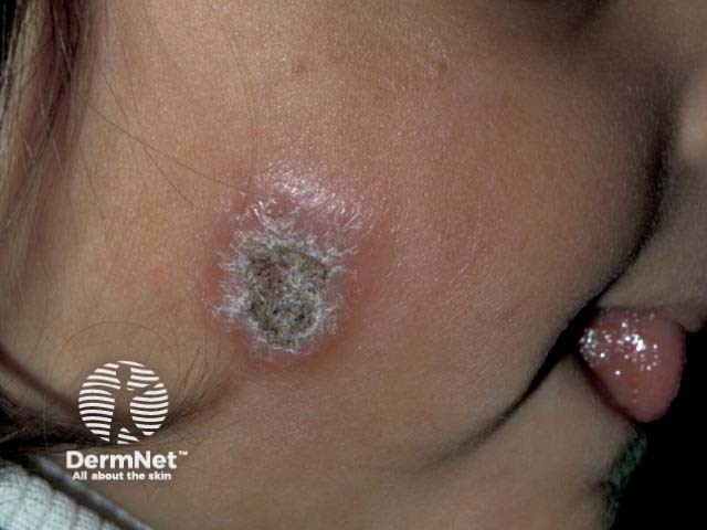
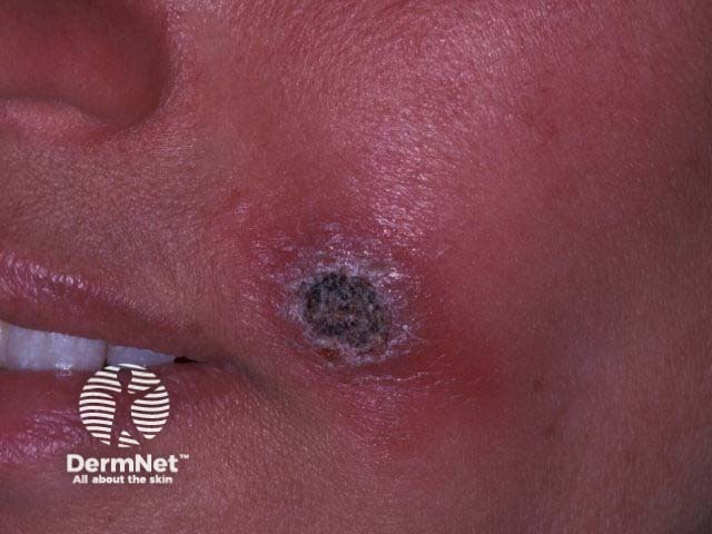
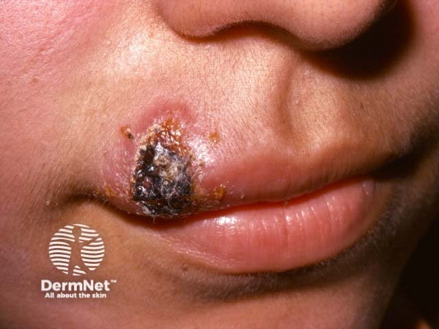
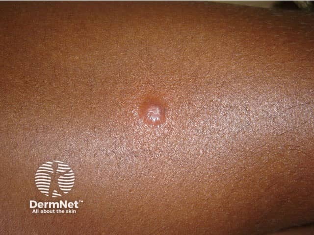
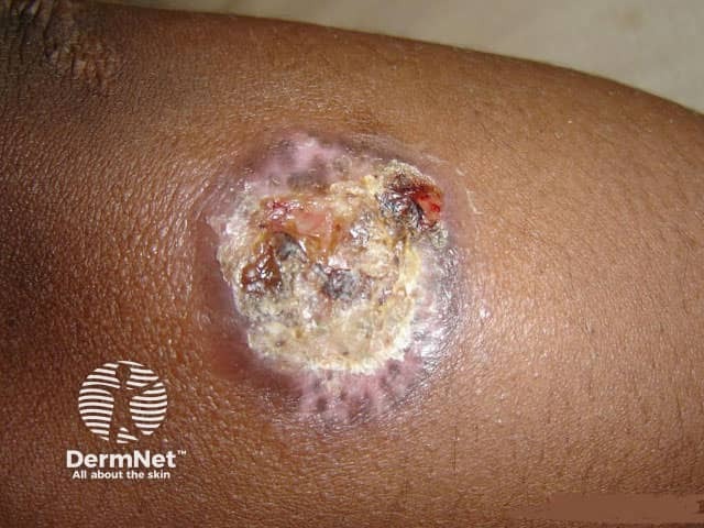
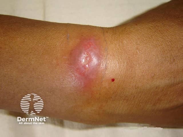
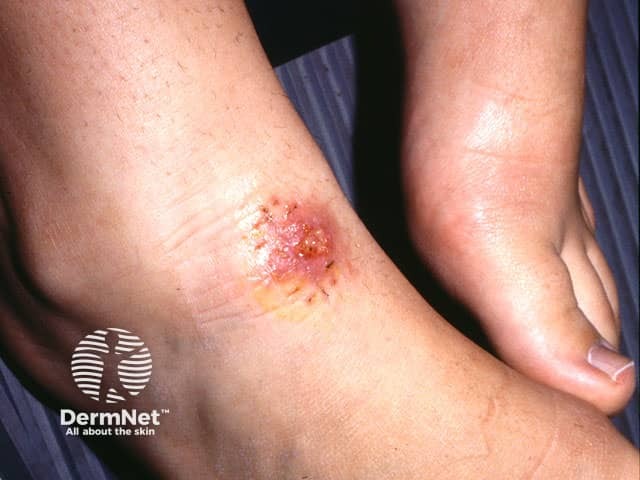
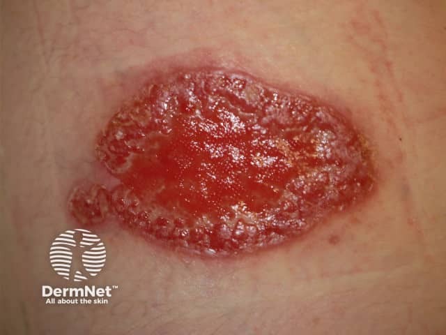
Cutaneous leishmaniasis typically occurs at the site of inoculation. The presentation and prognosis will vary depending on the species involved.
Synonyms: chiclero ulcer, uta, ulcera de Bauru, forest yaws, pian boi, bejuco.
Mucocutaneous leishmaniasis
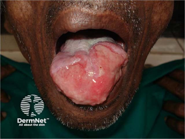
Mucocutaneous leishmaniasis is a destructive form of leishmaniasis, which is only seen with the American species of Leishmania (Viannia subspecies).
American mucocutaneous leishmaniasis:
Central and South America
L braziliensis, L guyanensis, L panamensis
Synonym: espundia.
Diffuse cutaneous leishmaniasis
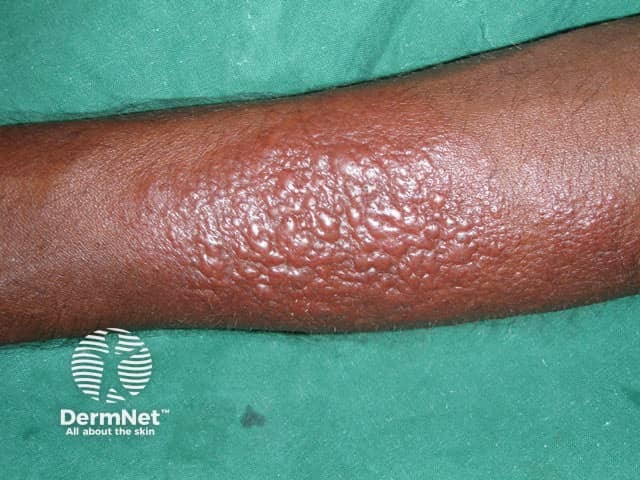
Diffuse cutaneous leishmaniasis is a rare presentation resulting from an anergic response to the parasite by the host.
Non-American diffuse cutaneous leishmaniasis:
Ethiopia, Kenya
L aethiopica.
American diffuse cutaneous leishmaniasis:
South America
L amazonensis.
Visceral leishmaniasis
Visceral leishmaniasis results from the involvement of the internal organs and is usually fatal if untreated. It is also known as kala-azar or Dumdum fever.
Non-American visceral leishmaniasis:
India, southern Europe, China, North Africa, Kenya
L donovani, L infantum, L tropica.
American visceral leishmaniasis:
Central and South America
L chagasi.
Post-kala-azar dermal leishmaniasis
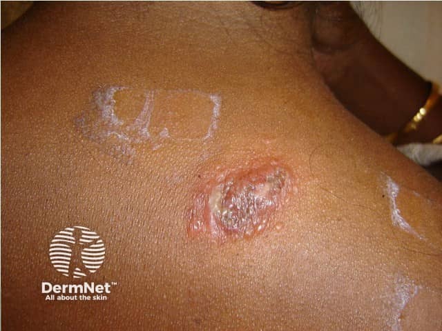
Post-kala-azar dermal leishmaniasis is a form of cutaneous leishmaniasis that can occur months to years after treatment of visceral leishmaniasis.
Non-American post-kala-azar dermal leishmaniasis:
Sudan, India
L donovani, L infantum, L tropica.
American post-kala-azar dermal leishmaniasis:
Central and South America
L chagasi.
Leishmaniasis recidivans
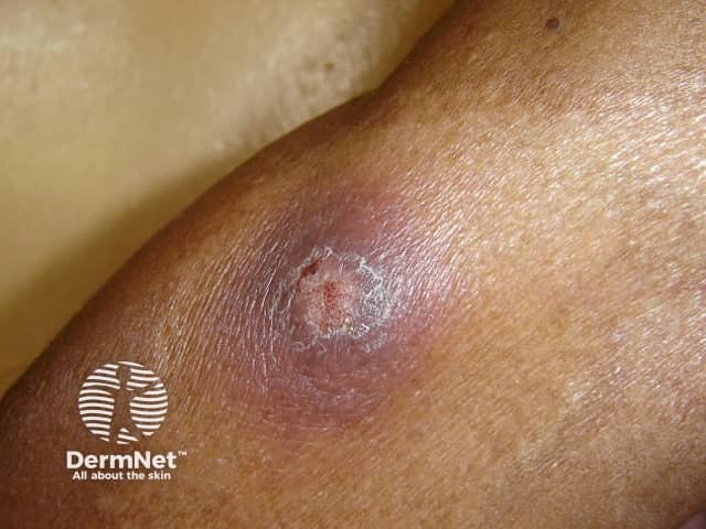
Leishmaniasis recidivans is a rare, cutaneous form of leishmaniasis, occurring in patients with a good cellular immune response. It is also known as lupoid leishmaniasis.
Non-American leishmaniasis recidivans:
Middle East, India, Southern Europe
L tropica.
American leishmaniasis recidivans:
Central and South America
L braziliensis.
What are the clinical features of leishmaniasis?
Cutaneous leishmaniasis
Cutaneous leishmaniasis is the most common form of leishmaniasis
Solitary lesions are typical, but multiple lesions do occur
The initial lesion is a small red papule, which gradually enlarges up to 2 cm in diameter
Central ulceration is typical
Ulcers can be moist and exude pus or dry with a crusted scab
Sores usually appear on exposed areas of the skin, especially the face and extremities
The incubation time between an infected sandfly bite and lesion development is ranges from 2 weeks to 6 months
Lesions are usually painless, and most resolve spontaneously often leaving residual atrophic scarring
Time to resolution varies between 2 months to more than a year
Sporotrichoid spread with lymphocutaneous nodules may occur
Chronic disease can occur, and there is a risk of dissemination in immunodeficient patients
Mucocutaneous leishmaniasis
Mucocutaneous leishmaniasis typically occurs after spontaneous resolution or local treatment of the primary cutaneous lesion
May develop within months or after many years
The lifetime risk of developing mucocutaneous leishmaniasis after a cutaneous lesion caused by L braziliensis is around 5%
Lesions usually affect the mucous membranes of the nose and mouth, but the mucosal surfaces of the eyes and genital tract can also be involved
Without treatment, ulceration of the mucosal surfaces and destruction of the underlying tissue can occur
Mucosal lesions are often painful and become secondarily infected, sometimes leading to sepsis
Can result in extreme disfigurement
Diffuse cutaneous leishmaniasis
Diffuse cutaneous leishmaniasis is a specific disease entity; sometimes the term is incorrectly used to describe disseminated or multiple cutaneous leishmaniasis
Results from an anergic response to the infection due to reduced cell-mediated immunity
Following the primary cutaneous leishmaniasis lesion, non-ulcerative nodules and plaques develop
Lesions may be numerous and may extend over the whole body
Follows a chronic relapsing or progressive course
Often difficult to treat
Visceral leishmaniasis
Visceral leishmaniasis affects internal organs including the spleen, liver and lymph nodes
Signs and symptoms include fever, weight loss, lymphadenopathy, hepatomegaly, and massive splenomegaly
Laboratory tests may show pancytopenia and hypergammaglobulinaemia
Complications include gastrointestinal bleeding, peripheraloedema, acuterenal failure, and secondary bacterialinfections
Generalisedhyperpigmentation is a late feature of visceral leishmaniasis; the other name for it, kala-azar, comes from the Hindi for ‘black fever’
Visceral leishmaniasis may take different forms ranging from asymptomatic self-resolving disease to being fulminant and life-threatening
Post-kala-azar dermal leishmaniasis
Post-kala-azar is a cutaneous form of leishmaniasis resulting as a complication of visceral leishmaniasis
It occurs months to years after treatment of visceral leishmaniasis
The majority of cases occur in Sudan (up to 50%) and India (up to 10%)
Lesions are either induratedpapules and nodules or areas of macularhypopigmentation
Most commonly affects the face, trunk and extremities
Oral mucosa and genital area may also be involved
Prognosis and treatment varies with location: in over 80% of Sudanese patients, the post-kala-azar dermal leishmaniasis lesions will resolve spontaneously within a year, whereas in India the rate of spontaneous resolution is much lower, so systemic therapy is used earlier
Leishmaniasis recidivans
Leishmaniasis recidivans occurs in patients with a good cellular immune response
Spontaneous resolution of the primary cutaneous lesion is followed by the development of new lesions around the edge of the primary scar
The lesions typically ulcerate then heal
The cycle continues with a chronic recurrent course, usually over decades
Cutaneous leishmaniasis in Sri Lanka
How is cutaneous leishmaniasis diagnosed?
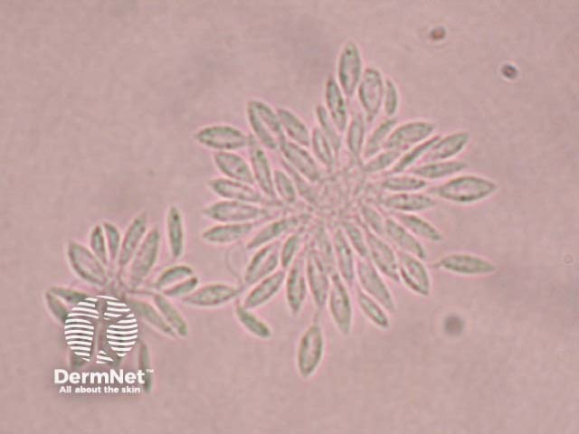
Diagnosis of cutaneous leishmaniasis is usually based on the history and clinical appearance of the lesion. A comprehensive travel history, including historical travel due to the long incubation period, is important in non-endemic areas. The diagnosis can be confirmed by identifying the parasite on biopsy or split skin smear. Culture and PCR may also be used to confirm the diagnosis and identify the species of Leishmania, which is important when there is a risk of mucocutaneous leishmaniasis.
Serology is used to confirm the diagnosis in visceral leishmaniasis.
In over 70% of cases, a full-thickness skin biopsy can reveal the parasite. Histopathology is also used to establish mucocutaneous leishmaniasis and visceral leishmaniasis. Complete blood counts and liver function tests should also be performed in visceral leishmaniasis.
New World leishmaniasis
Ulcer
Ulcer
What is the differential diagnosis for leishmaniasis?
The variety of clinical manifestations of cutaneous leishmaniasis results in a wide range of differential diagnoses:
In cutaneous leishmaniasis, treatment options differ depending on whether the lesion/s is considered simple or complex.
In non-American cutaneous leishmaniasis, lesions are considered complex if they are larger than 4 cm, multiple, associated with lymphatic spread, persistent (> 6 months), in immunosuppressed individuals, situated over joints, or in cosmetically sensitive areas.
All cases of American leishmaniasis should be considered complex because of the risk of mucocutaneous leishmaniasis with the Viannia subspecies.
Treatment options for cutaneous leishmaniasis lesions include:
Systemic antimonials (intravenous or intramuscular)
Sodium stibogluconate
Meglumine antimoniate.
Most cases of simple cutaneous leishmaniasis will resolve spontaneously without treatment, but this may take many months and can result in scarring.
Systemic antimonials are the mainstay of treatment for complex cutaneous leishmaniasis lesions, mucocutaneous leishmaniasis, and visceral leishmaniasis. They cannot be given orally, and the length of treatment may be up to 28 days for mucosal lesions. Treatment requires hospital admission, and there is a risk of side effects, including cardiotoxicity.
Secondary wound infections should be treated with appropriate antimicrobial therapy.
Surgical treatments and vaporising laser treatments are also used for appropriate lesions.
Refer to guidelines on the management of leishmaniasis published by the Infectious Diseases Society of America (IDSA) and the American Society of Tropical Medicine and Hygiene (ASTMH) (December 2016).
Prevention of insect bites
Infection can be prevented by avoidance of sandfly bites. Because there are currently no vaccines or drugs for preventing infection, travellers to areas where leishmaniasis is prevalent should decrease their risk of being bitten by adhering to the following precautionary measures.
Avoid outdoor activities, especially at dusk and dawn when sandflies are the most active.
Wear long-sleeved shirts, long pants, and socks — tuck shirt into pants.
Apply insect repellent on exposed skin and under the ends of sleeves and pant legs. The most effective repellents are those that contain the chemical DEET (N, N-diethyl-meta toluamide).
Spray clothing, living and sleeping areas (including bed net) with permethrin-containing insecticides.
References
Book: Textbook of Dermatology. Ed Rook A, Wilkinson DS, Ebling FJB, Champion RH, Burton JL. Fourth edition. Blackwell Scientific Publications.
Alvar J, Velez ID, Bern C, et al. Leishmaniasis worldwide and global estimates of its incidence. PLoS One 2012;7(5):e35671. DOI: 10.1371/journal.pone.0035671. PubMed.
Gonzalez U, Pinart M, Rengifo-Pardo M, Macaya A, Alvar J, Tweed JA. Interventions for American cutaneous and mucocutaneous leishmaniasis. Cochrane Database Syst Rev 2009; CD004834. DOI: 10.1002/14651858.CD004834.pub2. Review. PubMed.
Gonzalez U, Pinart M, Reveiz L. Alvar J. Interventions for Old World cutaneous leishmaniasis. Cochrane Database Syst Rev 2008; CD005067. DOI: 10.1002/14651858.CD005067.pub3. Review. PubMed.
Hashiguchi Y, Gomez EL, Kato H, Martini LR, Velez LN, Uezato H. Diffuse and disseminated cutaneous leishmaniasis: clinical cases experienced in Ecuador and a brief review. Trop Med Health 2016;44(2):DOI: 10.1186/s41182-016-0002-0. PubMed.