Authors: Ko Jin Quek, Junior Medical Officer, South-Western Sydney Local Health District, Sydney, NSW, Australia; Dr Monisha Gupta, Dermatologist, Sydney, Australia. DermNet Editor in Chief: Adjunct A/Prof Amanda Oakley, Dermatologist, Hamilton, New Zealand. Copy edited by Gus Mitchell. January 2020.
The term acquired dermal macular hyperpigmentation is useful because the three conditions overlap. The subtypes may manifest in the same patient, and they lack clear-cut clinical and histological differences.
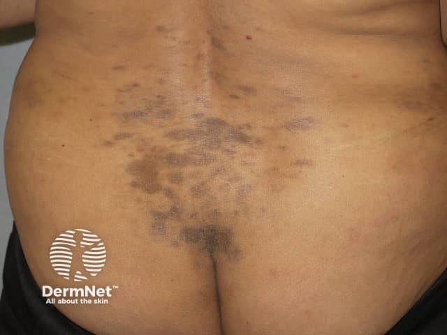
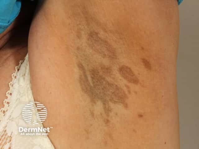
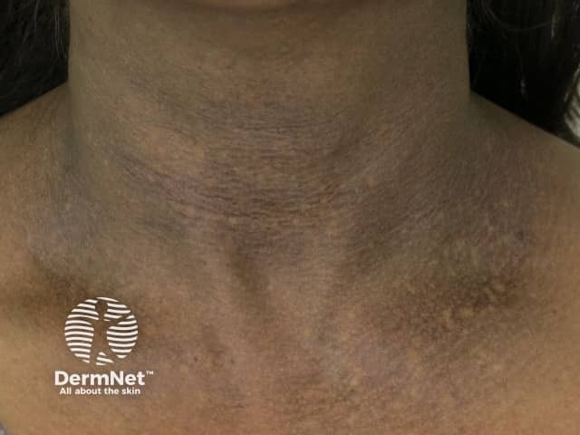
Who gets acquired dermal macular hyperpigmentation?
The epidemiology of acquired dermal macular hyperpigmentation varies depending on the underlying condition.
Erythema dyschromicum perstans can affect any age, sex, and ethnicity, and tends to affect darker-skinned patients. Asian, Middle-Eastern, and Latin American women between the 2nd and 4th decades of life are the most frequently affected. Whereas idiopathic eruptive macular hyperpigmentation develops in the first two decades of life. Lichen planus pigmentosus affects middle-aged people of South Asian and African/Middle Eastern descent.
What causes acquired dermal macular hyperpigmentation?
The exact cause of acquired dermal macular hyperpigmentation is unknown.
Proposed theories for the pathogenesis of acquired dermal macular hyperpigmentation include:
Geneticsusceptibility
Exposure to ultraviolet (UV) radiation
Contact reaction to a chemical (up to 36% of patients were reported to have a positive patch test reaction to hair dye), although this would now be termed pigmented contact dermatitis
The pigmentation in acquired dermal macular hyperpigmentation is often due to persistentmelanophages in the dermis. The reason the melanophages do not clear as usually occurs is unknown.
What are the clinical features of acquired dermal macular hyperpigmentation?
Acquired dermal macular hyperpigmentation presents with blue, brown, slate grey or brownish-black macules, which can change in size and morphology over time.
Erythema dyschromicum perstans affects the trunk and sun-protected locations, typically with an erythematous margin initially.
Large macules greater than 5 cm in diameter are seen in lichen planus pigmentosus and erythema dyschromicum perstans.
Small macules 0.5—2 cm in size occur in idiopathic eruptive macular hyperpigmentation and lichen planus pigmentosus.
Lichen planus pigmentosus is only rarely reported in patients with current or past classic lichen planus.
Acquired dermal macular hyperpigmentation is generally asymptomatic, although lichen planus pigmentosus is sometimes pruritic in its early phases.
What are the complications of acquired dermal macular hyperpigmentation?
No detrimental serious long-term medical complications arise from acquired dermal macular hyperpigmentation. However, it impacts on the quality of life in people with skin of colour due to cosmetic visibility and slow resolution.
How is acquired dermal macular hyperpigmentation diagnosed?
The diagnosis of acquired dermal macular hyperpigmentation and its subtypes are based on the cutaneous features and detailed history.
A detailed history is required to exclude medications, virus infection, and inflammatory skin conditions.
Patch testing may be indicated to exclude allergic contact dermatitis if there is a possible contact factor.
Dermoscopy is useful in the diagnosis and monitoring of acquired dermal macular hyperpigmentation. Findings may include:
Pigmented dots, globules and diffuse areas that spare eccrine and hairfollicle openings
Prominent normal pseudoreticular pigmentary network
Telangiectasia
Owl's eye structures.
Histological features of acquired dermal macular hyperpigmentation on skin biopsy may include and help to determine the diagnosis:
Melanin and melanophages in the dermis
Lichenoid changes with an interface dermatitis.
What is the differential diagnosis for acquired dermal macular hyperpigmentation?
A variety of other skin conditions appear similar to acquired dermal macular hyperpigmentation, such as:
What is the treatment for acquired dermal macular hyperpigmentation?
The treatment of acquired dermal macular hyperpigmentation depends on the subtype and its duration, but many therapies have been tried with little or no benefit.
In later stages or where there is no acute inflammation, treatment aims to reduce pigmentation; stringent sun protection is advised. Topical retinoids may reduce melanin that is retained within the epidermis.
What is the outcome for acquired dermal macular hyperpigmentation?
Idiopathic eruptive macular hyperpigmentation tends to resolve spontaneously within months to years. In contrast the pigmentation of lichen planus pigmentosus can persist for decades and erythema dyschromicum perstans tends to be chronic and progressive.
References
Sasidharanpillai S, Govindan A, Ajithkumar KY, et al. Histological evaluation of acquired dermal macular hyperpigmentation. Indian Dermatol Online J 2019; 10: 542–6. doi: 10.4103/idoj.IDOJ_426_18. PubMed Central
Udompanich S, Vachiramon V. Acquired macular pigmentation of unknown etiology. J Clin Aesthet Dermatol 2019; 12: 38–46. PubMed Central
Rodrigues M, Pandya AG, Bekkenk M, Parsad D, Kumarasinghe SP. Current understanding of lichen planus pigmentosus, erythema dyschromicum perstans (ashy dermatosis), and idiopathic eruptive macular pigmentation. Pigment Int 2019; 6: 4–8. doi: 10.4103/Pigmentinternational.Pigmentinternational. Journal
Chang SE, Kim HW, et al. Clinical and histological aspect of erythema dyschromicum perstans in Korea: a review of 68 cases. J Dermatol 2015; 42: 1053–7. doi: 10.1111/1346-8138.13002. PubMed
Kumarasinghe SPW, Pandya A, Chandran V, et al. A global consensus statement on ashy dermatosis, erythema dyschromicum perstans, lichen planus pigmentosus, idiopathic eruptive macular pigmentation, and Riehl's melanosis. Int J Dermatol 2019; 58: 263–72. doi: 10.1111/ijd.14189. PubMed
Bishnoi A, Vinay K, Arshdeep, et al. Contact sensitization to hair colours in acquired dermal macular hyperpigmentation: results from a patch and photo-patch test study of 108 patients. J Eur Acad Dermatol Venereol 2019; 33: 1349–57. doi: 10.1111/jdv.15576. PubMed
Vinay K, Bishnoi A, Parsad D, Saikia UN, Sendhil Kumaran M. Dermatoscopic evaluation and histopathological correlation of acquired dermal macular hyperpigmentation. Int J Dermatol 2017; 56: 1395–9. doi: 10.1111/ijd.13782. PubMed
Kanwar AJ, Parsad D. Colchicine in the treatment of Lichen Planus Pigmentosus, XXIst International Pigment Cell Conference, Bordeaux, France, 2011.
Al-Mutairi N, El-Khalawany M. Clinicopathological characteristics of lichen planus pigmentosus and its response to tacrolimus ointment: an open label, non-randomized, prospective study. J Eur Acad Dermatol Venereol 2010; 24: 535–40. doi: 10.1111/j.1468-3083.2009.03460.x. PubMed
Bahadir S, Cobanoglu U, Cimsit G, Yayli S, Alpay K. Erythema dyschromicum perstans: response to dapsone therapy. Int J Dermatol 2004; 43: 220-2. doi: 10.1111/j.1365-4632.2004.01984.x. PubMed
Kontochristopoulos G, Stavropoulos P, Panteleos D, Aroni K. Erythema dyschromicum perstans: response to dapsone therapy. Int J Dermatol 1998; 37: 796–8. doi: 10.1046/j.1365-4362.1998.00522.x.