Sporotrichoid lymphocutaneous infection is characterised by the appearance of subcutaneousnodules that progress along dermal and lymphatic vessels. It is also called nodularlymphangitis.
The clinical presentation is often described as sporotrichoid spread. The name 'sporotrichoid' is because the most common infection is sporotrichosis.
Deep fungal infection
Nocardia
Nocardia
What does sporotrichoid spread look like?
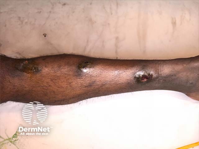
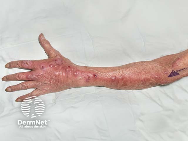
Sporotrichoid spread is mostly observed on an upper limb. The first lesion starts distally, for example, on the hand, wrist or forearm. Subsequent lesions arise proximally, that is, further up the arm, in an irregular, roughly linear, distribution.
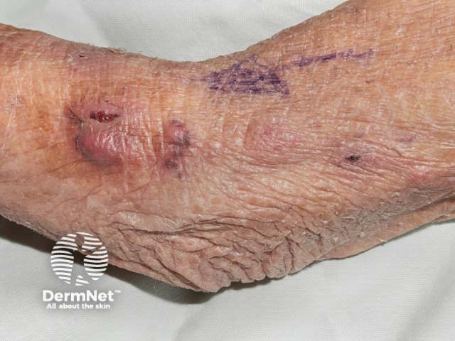
Each lesion is an inflamed, irregular, firm nodule, which may suppurate or ulcerate.
What is the cause of sporotrichoid lymphocutaneous infection?
Sporotrichoid lymphocutaneous infection is usually due to an uncommon infection transmitted by primaryinoculation through a minor injury or insect bite:
Skin biopsy for histopathology (which will show granulomas and abscess formation) and special stains to locate and identify the organisms.
How is sporotrichoid lymphocutaneous infection treated?
Treatment depends on the cause, so it is very important to identify the organism before starting a course of antibiotics or oral antifungal medicine. For example:
Leishmania may be treated by stibogluconate or amphotericin.
Other conditions with sporotrichoid spread
Lesions of cutaneous sarcoidosis are also occasionally noted to have sporotrichoid spread.
In-transit metastases are sometimes considered in the differential diagnosis of lymphocutaneous infection, where a secondary tumour – usually melanoma – arises in the skin between the excision site of the primary melanoma and its nearest lymph node.
References
Tobin E, Jih WW. Sporotrichoid Lymphocutaneous Infections: Etiology, Diagnosis and Therapy. Am Fam Physician 2001;63:326–32. PubMed