
ADVERTISEMENT
You do not have any notes added to this page yet
Introduction
Demographics
Causes
Clinical features
Complications
Diagnosis
Differential diagnoses
Treatment
Outcome
A xanthoma is a skin lesion caused by the accumulation of fat in macrophages in the skin. Less commonly, a xanthoma will occur in a subcutaneous layer.
Xanthomas are usually a skin sign of disorders of lipid metabolism (dyslipidaemias) or occur in histiocytosis; the former is the focus of this page. Xanthomas typically affect adults, although children with familial hypercholesterolaemia may present with xanthomas. The race and sex distributions depend on the underlying cause.
Dyslipidaemias causing xanthomas are classified as primary or secondary.
Primary dyslipidaemias associated with xanthomas can include:
Secondary dyslipidaemias associated with xanthomas include:
Diffuse plane xanthomatosis is associated with paraproteinaemia. Xanthoma disseminatum is a rare form of histiocytosis.
There are multiple types of xanthomas, and they are classified by their clinical presentation.
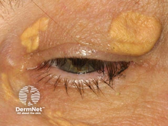
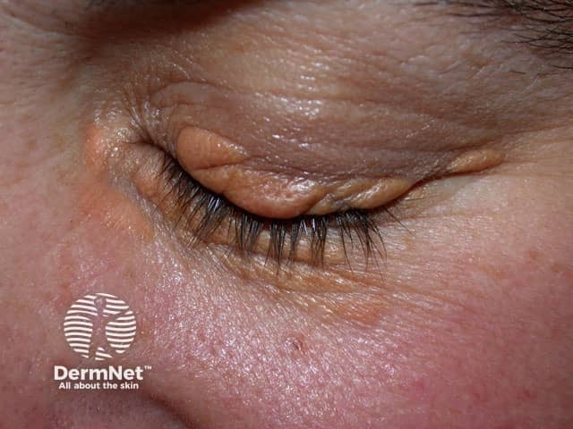
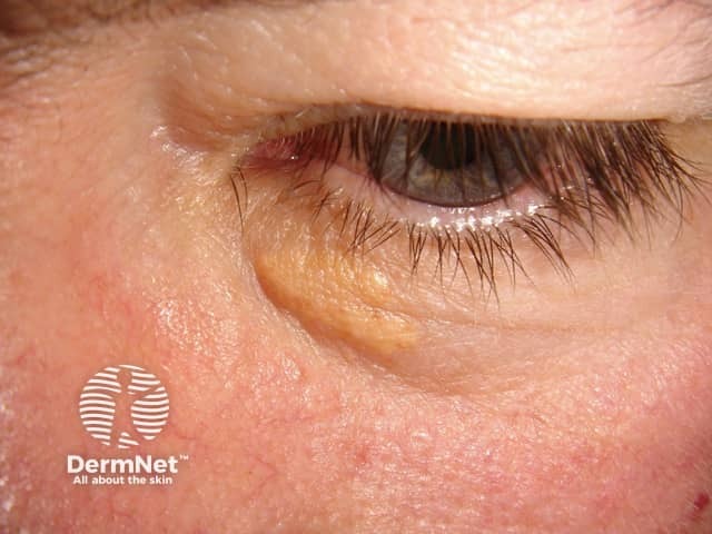
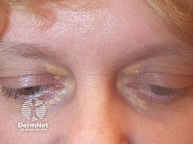
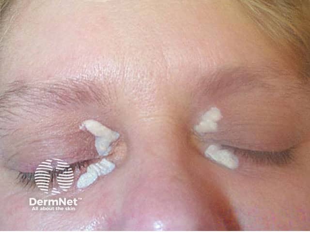
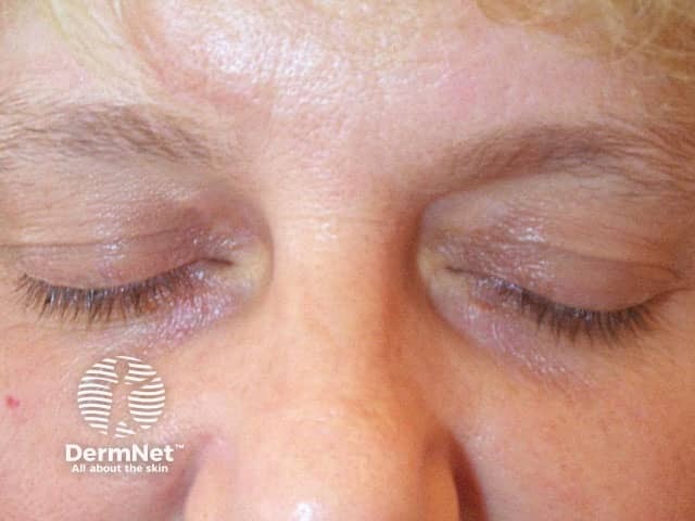
Xanthelasma (also known as xanthelasma palpebrarum or xanthelasma palpebrum), is the most common form of xanthoma. It is a type of plane xanthoma usually located around the medial canthus of the upper eyelid. Upper and lower eyelids can be affected symmetrically.
Xanthelasma:
See more images of xanthelasma.
Plane xanthomas (planar xanthoma, plane xanthomatosis) are soft, yellow macules or patches that can occur anywhere on the body. Involvement of the webspace between the fingers and toes is pathognomonic for homozygous familial hypercholesterolaemia. Plane xanthomas may also be associated with Type III hyperlipoproteinaemia, and need to be distinguished from diffuse plane xanthomatosis.
Palmar xanthoma (xanthoma striata palmaris) present as a yellow-orange accentuation of the palmar and wrist creases. This is diagnostic for Type III hyperlipoproteinaemia.
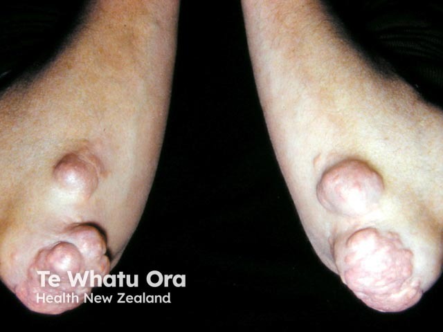
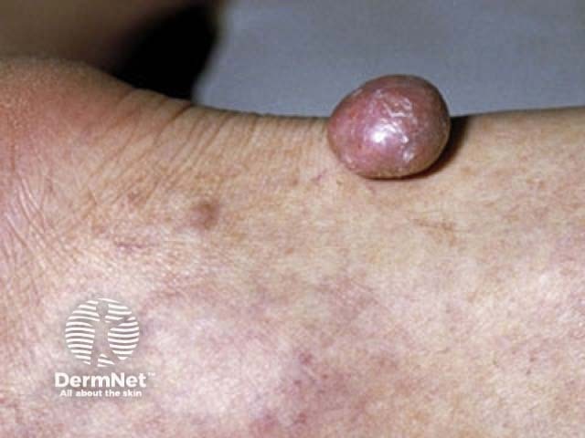
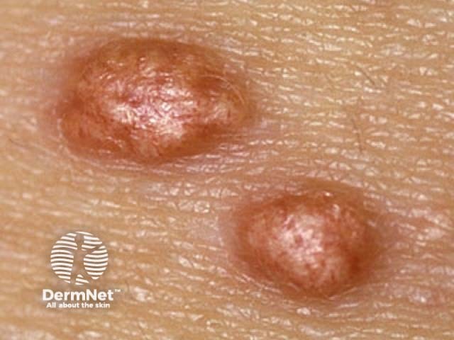
Tuberous xanthomas are firm, painless, red-yellow nodules that develop over pressure areas such as the knees, elbows, and heels. They may join together to form large multilobulated masses. Tuberous xanthomas are often associated with Type III hyperlipoproteinaemia.
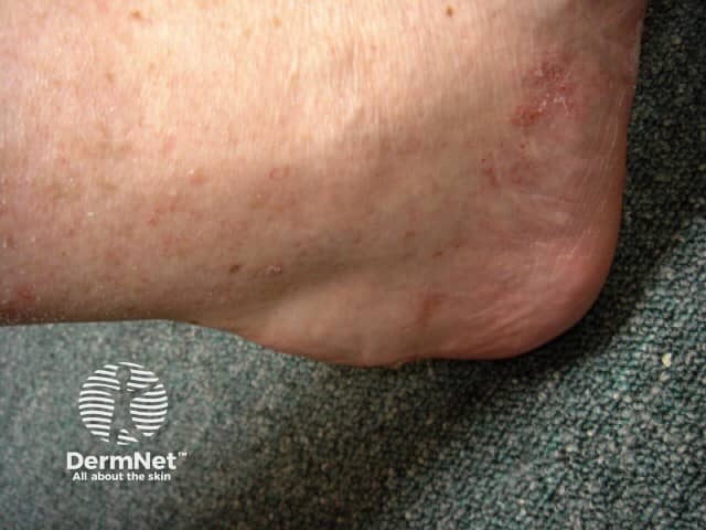
Tendon xanthomas (tendinous xanthoma) are slowly enlarging subcutaneous nodules usually found attached to the Achilles tendon or tendons over the knuckles. They are smooth, firm to palpation, and mobile. The overlying skin colour is normal. Tendon xanthomas are most commonly associated with familial hypercholesterolaemia, but can also be seen in cerebrotendinous xanthomatosis and sitosterolaemia.
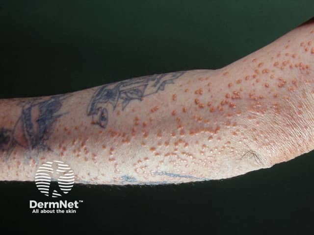
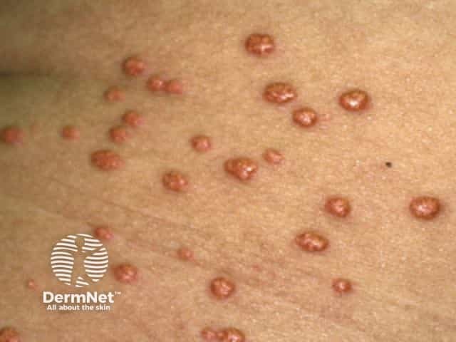
Eruptive xanthomas typically present as crops of 2–5 mm yellow papules with a red rim over extensor surfaces such as the buttocks or shoulders, but can be widespread including inside the mouth. The papules may be tender and are usually itchy. They may demonstrate köbnerisation. Eruptive xanthomas are due to hypertriglyceridaemia (triglyceride >11.2 mmol/L) of any cause.
Verruciform xanthoma is a rare entity not associated with dyslipidaemias. It most commonly affects the mouth, where it is found as a solitary, asymptomatic lesion on the gingiva and is associated with chronic graft-versus-host disease. On the genitalia, it has been called a Vegas (verruciform genital-associated) xanthoma and presents as a yellow-brown or red verrucous plaque. The lipid found in the foamy macrophages is thought to derive from keratinocytes, and it is possibly a reaction to chronic irritation.
Xanthoma is usually a sign skin of a dyslipidaemia and therefore the complications are those of the underlying condition, such as pancreatitis or cardiovascular disease. Xanthelasma appears to be an independent predictor of ischaemic heart disease, separate from any associated dyslipidaemia.
Xanthoma is often a clinical diagnosis made in the setting of a dyslipidaemia. Skin biopsy may be required and shows the characteristic lipid-filled macrophages in the dermis (see Eruptive xanthoma pathology).
Investigations are required to determine the associated condition and may include:
Xanthomas may need to be distinguished from many other skin lesions depending on the clinical presentation.
Some xanthomas resolve with successful treatment of the underlying dyslipidaemia: tuberous, eruptive, plane, and palmar. Xanthelasma may improve if associated with hypercholesterolaemia and this can be successfully treated. However treatment of the lesions may be required for cosmetic reasons and can include topical trichloroacetic acid (see Chemical peels), electrodessication, cryotherapy, laser vaporisation (see Nd:YAG, Er:YAG, carbon dioxide laser), or excision. Verrucous xanthoma is surgically excised and rarely recurs.

The importance of diagnosing xanthomas is recognising the associated trigger and its complications. The prognosis therefore depends on the successful management of the underlying medical condition.
An AI summary will appear based on your search term using data from all of the topic pages across the entire DermNet site.
Show more