Author(s): Linda Mardiros, University of Ottawa, Canada; Hana Numan, Senior Medical Writer, DermNet (2022) Previous contributors: Hon A/Prof Amanda Oakley, Dermatologist (2016) Reviewing dermatologist: Dr Ian Coulson
Leukonychia can be classified by underlying pathology, its distribution, or how it develops.
Classification according to pathology
Leukonychia can be subdivided into true and apparent discolouration.
True leukonychia: discolouration due to abnormal nail plate keratinisation. The white nail will not be hidden by pressure application of the nail plate to the bed.
Apparent leukonychia: secondary to disease of the nail bed. This appearance disappears with pressure application on the nail.
Classification according to distribution
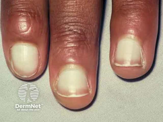
Leukonychia can be partial or total.
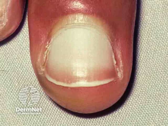
Total leukonychia: whitening of the entire nail plate.
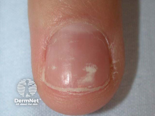
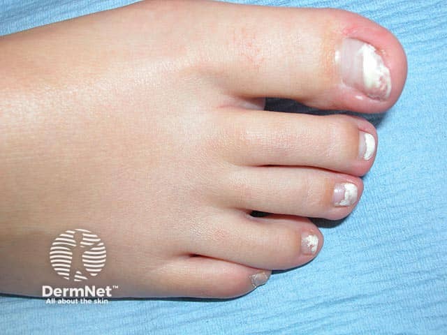
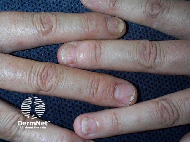
Partial leukonychia: 3 subtypes are described.
Punctate
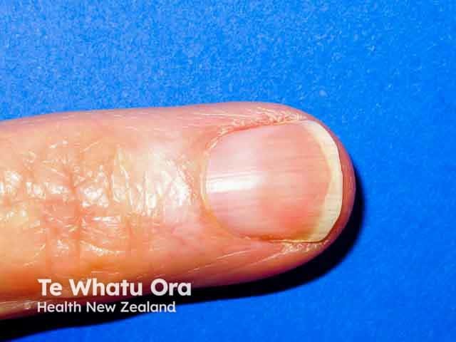
Longitudinal
Striate (see below).
Classification by development timeline
White nails can be acquired or congenital.
Congenital: familial leukonychia is more commonly inherited recessively, although dominant patterns are possible. This is due to a mutation in the phospholipase C delta-1 gene in which all nails appear milky and porcelain white.
Acquired: secondary to systemic disease. Important to note that congenital leukonychia may also be secondary to systemic disease (see below).
Who gets a white nail?
White nails can affect anyone of any gender, age or ethnicity. Its presence may warrant a work-up for systemic disease.
What causes a white nail?
Trauma
True leukonychia: partial or whole nail plate damage caused by injury to the nail plate or matrix. Keratin disruption with trapped air within the nail plate, resulting in reflection and lack of transparency.
Punctate leukonychia: occurs after nail biting, manicuring, knocks and bangs, and tight footwear use.
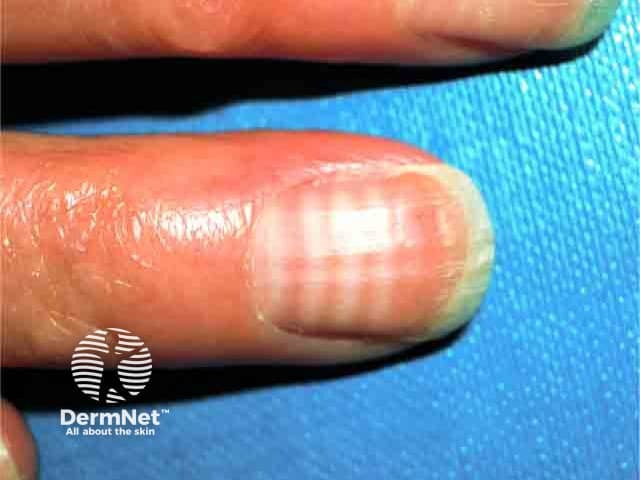
Striate leukonychia: also known as Mees lines or transverse leukonychia, may follow damage to the nail matrix; furrows and ridges may also appear.
Total leukonychia: can follow a more serious injury, often with detachment of the nail plate from the nail bed, and alteration to the nail contour.
Poisoning and drugs
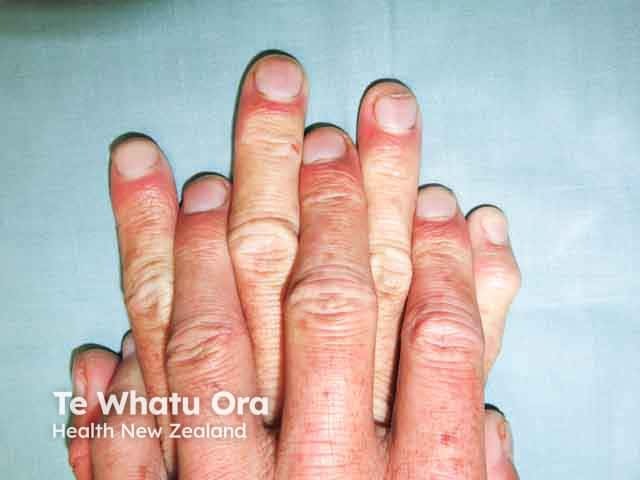
Mees line, Lindsay nails, Muehrcke lines (see below), and punctate leukonychia may be associated with:
One or more white horizontal bands across the entire nail in parallel with the lunula.
Patients with multiple true leukonychia warrant a thorough history, physical examination, and medication review to exclude a toxic or systemic etiology. This is also true of leukonychia extending the full width of the nail plate.
Treatment ultimately depends on the presence of any underlying cause.
There is no treatment for trauma-related leukonychia. Punctate lesions will disappear as the nail follows its natural growth pattern (around 6 to 9 months for a fingernail).
How do you prevent a white nail?
Avoidance of trauma-induced leukonychia will help prevent development of white nails.
Avoiding contact with irritating substances, and wearing appropriate protective equipment if contact is required.
Avoiding excessive use of nail polish or excessive mechanical force with false nail application/removal.
Minimising picking and biting nails.
Wearing appropriate shoes to prevent excessive pressure on toes.
Leukonychia due to minor trauma or medication may completely resolve over a few months. In other cases, the white nail plate may remain permanently or demonstrate recurrence.
Bibliography
Iorizzo M, Starace M, Pasch MC. Leukonychia: What Can White Nails Tell Us?. Am J Clin Dermatol. 2022;23(2):177–93. doi:10.1007/s40257-022-00671-6. Journal
Pitukweerakul S, Pilla S. Terry's Nails and Lindsay's Nails: Two Nail Abnormalities in Chronic Systemic Diseases. J Gen Intern Med. 2016;31(8):970. doi:10.1007/s11606-016-3628-z. Journal
Richert B, Caucanas M, André J. Diagnosis using nail matrix. Dermatol Clin. 2015;33(2):243–55. doi:10.1016/j.det.2014.12.005. Journal