Author: Prof. Balachandra Ankad, Dermatologist, S Nijalingappa Medical College, Karnataka, India. DermNet Editor in Chief: Adjunct A/Prof Amanda Oakley, Dermatologist, Hamilton, New Zealand. October 2019.
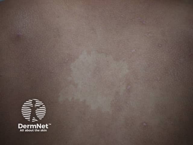
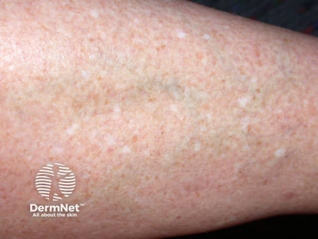
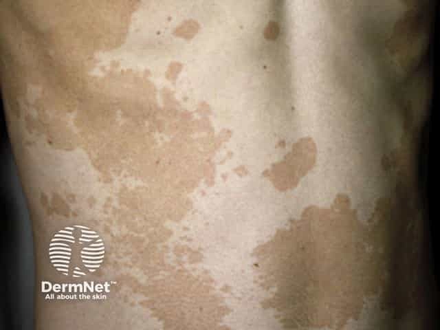
Vitiligo is a common acquired depigmentation of the skin due to autoimmune destruction of melanocytes. It is characterised by well-circumscribed chalky-white macules and patches. Hairs in the involved skin may be normal or white.
What are the clinical features of vitiligo?
The clinical manifestations of vitiligo include depigmented macules and patches on the skin, mucous membranes, and hair. White hairs in the involved area are associated with a poor prognosis.
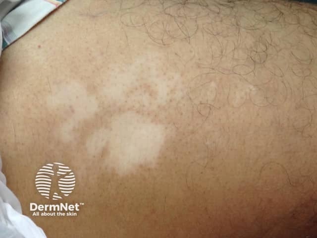
Vitiligo on the gluteal area
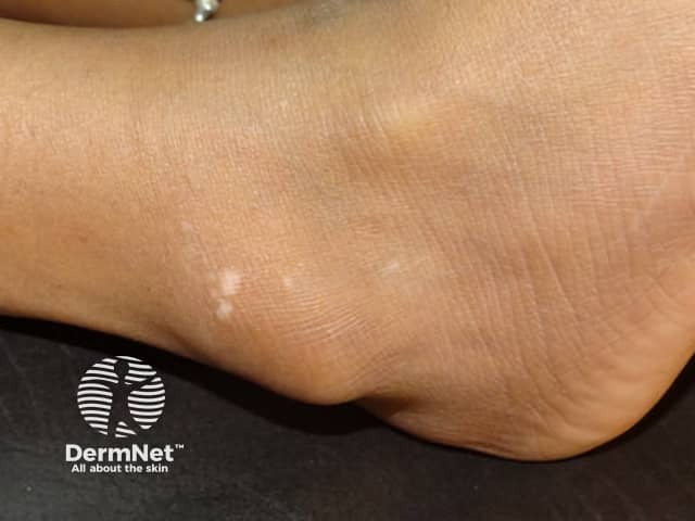
Vitiligo of the ankle
Vitiligo
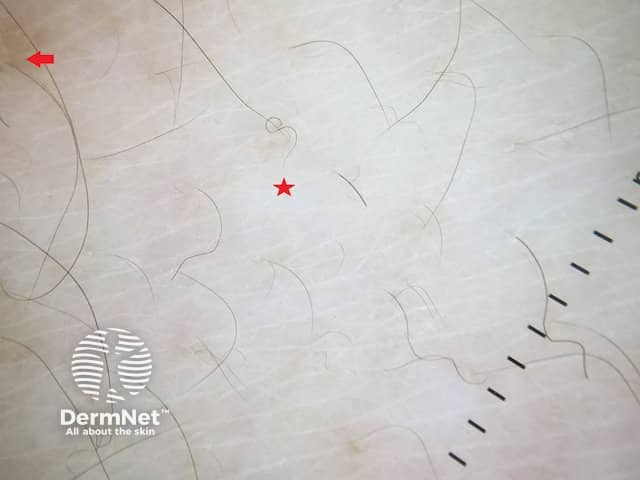
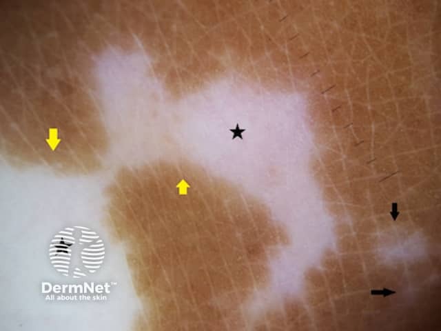
What are the dermoscopic features of vitiligo?
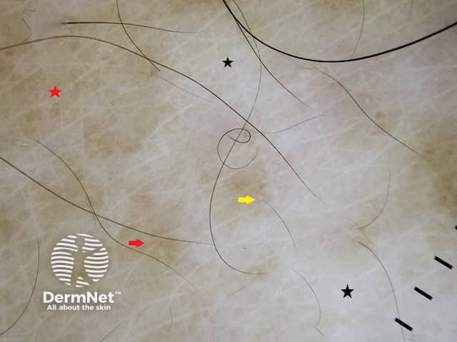
White structureless areas (absence of pigment network) and reduced pigment network. White structureless areas appear to ‘glow’ due to total loss of melanocytes in the epidermis
Perilesional and perifollicularhyperpigmentation and reversed pigment network
Stable vitiligo is characterised by a sharp border, white structureless areas, reduced pigment network, and perilesional and perifollicular hyperpigmentation, whereas active disease demonstrates a starburst pattern, tapioca sago pattern (satellite lesions), micro-koebnerisation and comet-tail appearance.
What is the dermoscopic differential diagnosis for vitiligo?
Achromic naevusdermoscopy: white structureless areas with a faint pigment network in the background. Glow is absent due to the presence of the pigment network. White areas may extend peripherally. [see Dermoscopy of achromic naevus]
Idiopathic guttate hypomelanosis dermoscopy: shows a similar glow but unlike vitiligo, the white area is clearly demarcated from normal skin. Guttatehypomelanosis has amoeboid (pseudopod-like border), petaloid, feathery and nebuloid (gradually fading border) morphological variants. [see Idiopathic guttate hypomelanosis dermoscopy]
Pityriasis alba dermoscopy: white structureless areas without a glow, a faint pigment network, and superficial white scales.
Pityriasis versicolor dermoscopy: also white structureless areas without a glow, pigment network and white scales. Scales are prominent in the skin lines and are separated into lines when the lesion is stretched. [see Dermoscopy of pityriasis versicolor]
Achromic naevus
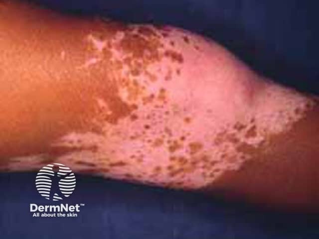
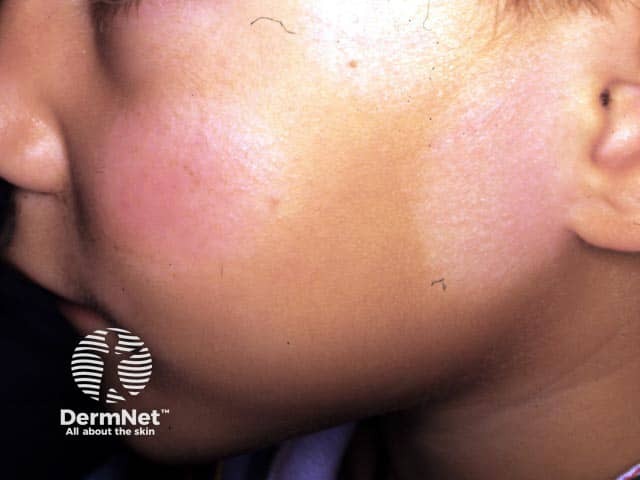
Pityriasis alba on the cheeks
Pityriasis versicolor
What is the histological explanation of the dermoscopic features of vitiligo?
The histology of vitiligo shows a normal epidermis with loss of melanocytes. A normal reticulate pigment network is due to melanocytes in the rete ridges. In vitiligo, destruction of melanocytes results in loss of the pigment network resulting in the dermoscopic glow.
References
Ezzedine K, Lim HW, Suzuki T, et al. Revised classification/nomenclature of vitiligo and related issues: the Vitiligo Global Issues Consensus Conference. Pigment Cell Melanoma Res. 2012;25(3):E1–13. doi: 10.1111/j.1755-148X.2012.00997.x. PubMed PMID: 22417114; PubMed Central PMCID: PMC3511780.
Kumar Jha A, Sonthalia S, Lallas A, Chaudhary RKP. Dermoscopy in vitiligo: diagnosis and beyond. Int J Dermatol. 2018;57(1):50–4. doi:10.1111/ijd.13795. Epub 2017 Oct 26. PubMed PMID: 29076154.
Nirmal B, Antonisamy B, Peter CVD, et al. Cross-sectional study of dermatoscopic findings in relation to activity in vitiligo: BPLeFoSK Criteria for Stability. J Cutan Aesthet Surg. 2019;12(1):36–41. doi: 10.4103/JCAS.JCAS_75_18. PubMed PMID: 31057267; PubMed Central PMCID: PMC6484572.