Trichofolliculoma presents as a small, solitary flesh-coloured or whitish nodule that occurs most often on the face around the nose region. Centrally, there is a pore which sometimes may contain numerous hairs.
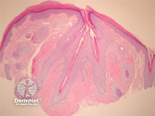
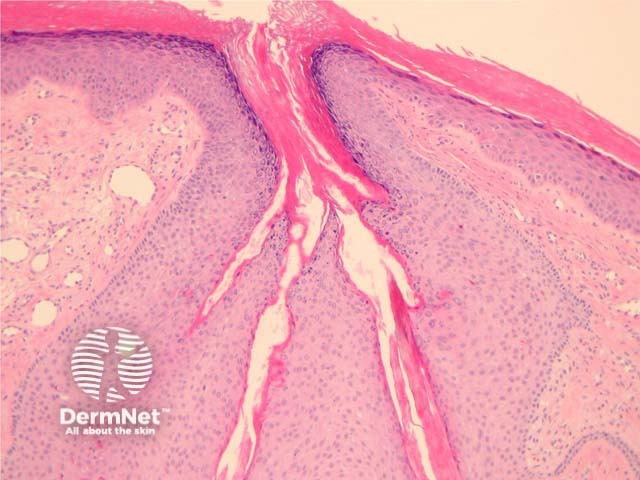
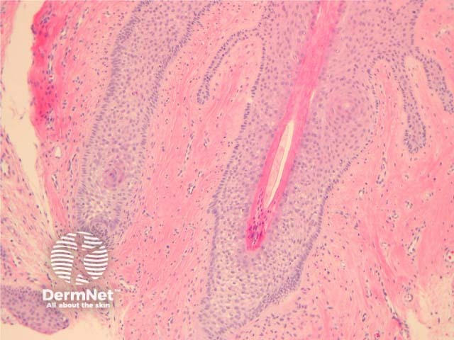
Histology of trichofolliculoma
In trichofolliculoma, sections show numerous small follicles radiating from a central larger follicle. The hairs are surrounded by a well circumscribed dense stroma. (figures 1–3). The surrounding hairs are generally very small (vellus). Intermixed Merkel cells and sebocytes may be seen. A sebaceous variant has been described (sebaceous trichofolliculoma).
As with all folliculartumours, there may be numerous mitoses/apoptotic figures within basaloid cell nests which are contributing to the production of a hair shaft – these should not be confused with a proliferatingmalignant process.
Figure 1
Figure 2
Figure 3
Special studies for trichofolliculoma
None are generally needed.
Differential diagnosis of trichofolliculoma
Basal cell carcinoma – BCC shows retraction between the tumour cells and the surrounding stroma. BCCs show more nuclearatypia, often an infiltrative growth pattern.
Trichodiscoma/fibrofolliculoma – Follicular epithelium surrounded by a perifollicular fibrous sheath. The epithelial strands are composed of thin cords of epithelial cells. These may be associated with Birt-Hogg-Dubé syndrome.
Neurofollicular hamartoma – These are thought to be a spindle cell rich form of trichodiscoma by some authorities. The spindle cells are often positive with S100 and CD34.
References
Pathology of the Skin (Fourth edition, 2012). McKee PH, J. Calonje JE, Granter SR
Misago N, Kimura T, Toda S, Mori T, Narisawa Y. A revaluation of trichofolliculoma: the histopathological and immunohistochemical features. Am J Dermatopathol. 2010 Feb;32(1):35–43. PubMed