Toxic epidermal necrolysis (TEN) is a severe cutaneous drug reaction characterised by a prodromal 'flu-like illness followed by the rapid appearance of a painful erythematous rash and desquamation of skin and mucous membranes. TEN is at the severe end of a spectrum with Stevens-Johnson syndrome defined by >30% body surface area skin detachment.
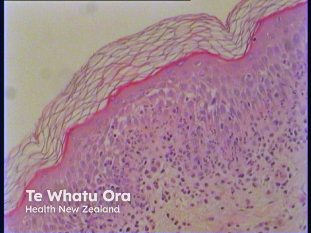
Histology of toxic epidermal necrolysis
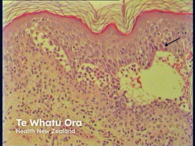
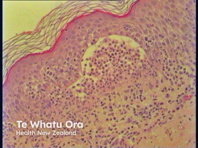
In TEN, there are subepidermalbullae (Figures 1–3) with widespreadepidermalnecrosis and subsequent separation or loss of the entire epidermis.Apoptotickeratinocytes may be seen at the periphery (Figure 2, arrow). There is minimal inflammatoryinfiltrate.
Figure 1
Figure 2
Figure 3
Differential diagnosis of toxic epidermal necrolysis
Erythema multiforme: Apoptotic keratinocytes with less prominent necrosis (may be seen at the centre of established lesions), inflammatory infiltrate is more prominent with lymphocyticperivascular infiltrate.
Phytophototoxic reaction: Marked oedema and subepidermal blistering with apoptotic keratinocytes and epidermal necrosis. Minimal inflammatory infiltrate. Clinical features will easily differentiate from TEN.
Chemotherapy-induced acral erythema: If severe can progress to subepidermal blistering with epidermal necrosis. Clinically distinct from TEN.
Differential diagnosis of cell-poor subepidermal blistering includes variants of epidermolysis bullosa, porphyria cutanea tarda, cell-poor type bullous pemphigoid, burns, suction blisters and some bullous drug reactions.