
ADVERTISEMENT
You do not have any notes added to this page yet
Introduction
Acute inflammatory reactions
Skin infections
Eczematous hypersensitivity reactions
Photo-aggravated reactions
Granulomatous reactions
Psoriasis
Lichenoid reactions
Pseudolymphomatous reactions
Skin cancer
Burns during magnetic resonance imaging
Removal of tattoos
Decorative tattooing has been practised for thousands of years. In primitive times it was used for embellishment, whilst in some customs and cultures tattooing represented a sign of distinction or social rank. This remains the case today with some cultures; however, it has also become popular with everyday people of western countries in the last 10–20 years.
With the rise in the number of people with tattoos in today's society, an increase in the number of tattoo-associated skin disorders can be expected. Reactions that may occur include acute inflammatory reactions, eczematous hypersensitivity reactions (allergic contact dermatitis), photo-aggravated reactions, granulomatous reactions, lichenoid reactions and pseudolymphomatous reactions.

An acute inflammatory reaction is in direct response to the piercing of the skin with needles impregnated with pigment dyes prepared from metal salts. There may be transient redness and swelling of the area that disappears within 2–3 weeks. It is an expected side effect of the tattooing process.
The koebner phenomenon describes the appearance of new lesions of an existing skin disease within a cutaneous injury. A tattoo can result in psoriasis, lichen planus or vitiligo by this mechanism.
Infection is not common after tattooing. The following skin infections have been reported however, emphasising the need to undergo the procedure in a clean environment using sterile equipment. Microorganisms have also been found in contaminated tattoo ink.
Transmission of serious blood-borne infections may occur.
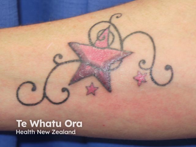
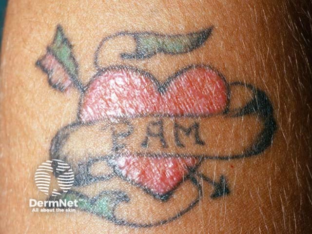
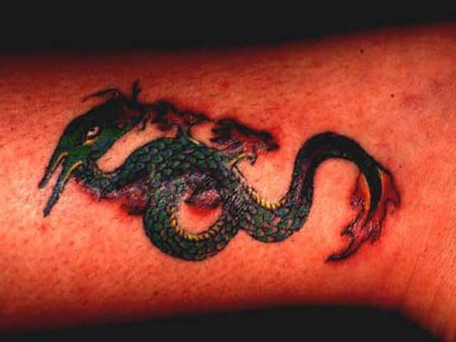
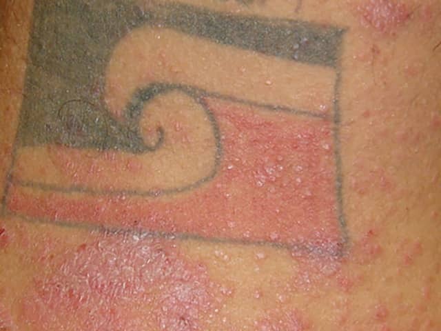
The two most common hypersensitivity reactions to tattoo pigments are allergic contact dermatitis and photoallergic dermatitis. The reaction usually appears as an inflamed red rash or may sometimes be scaly and flaky (exfoliative dermatitis). Red tattoo pigments cause the most reactions, particularly those made from mercury sulfide (cinnabar). Hypersensitivity reactions to pigments used to make black, blue, purple and green tattoos are much less common.
The components of tattoo ink are difficult to determine and undergo changes with time. There are no regulations for tattoo inks or colour additives, which contain inorganic pigments and carbon black, and/or organic pigments from various chemical classes. The table below lists some agents that have been used. There are many more.
Colour |
Composition |
|
|---|---|---|
Red |
|
|
Black |
|
|
Brown |
|
|
Blue |
|
|
Green |
|
|
Yellow |
|
|
Purple |
|
|
White |
|
|
Allergic contact dermatitis has also been reported in people with henna tattoos. Henna tattoos are non-permanent tattoos where henna dye is painted onto the skin with an artist's brush resulting in a brownish stain. Henna itself should be safe, but the dye is often mixed with paraphenylenediamine (PPD), a chemical substance that is well known for causing allergic reactions in people sensitive to it. In this case, the dye is black in colour, so-called black henna.
Yellow tattoos created from cadmium sulfide are at most risk of causing hypersensitivity reactions when they are exposed to sunlight. Swelling and redness develop around the tattoo site. This phototoxic reaction caused by cadmium sulfide can also occur in red tattoos, as trace amounts of cadmium are added to brighten red tattoo pigment.
The term granuloma refers to the particular kind of cells that cause the reaction. A foreign body reaction to pigment may cause raised red bumps at the site of the tattoo that are made up of epithelioid cells, lymphocytes and a few giant cells. Most commonly red, but also, green, blue and purple pigment tattoos, and UV-visible tattoos have been associated with granulomatous reactions. Granulomatous reactions in tattoos can also occur in patients with sarcoidosis.
Psoriasis has been reported in tattoos, often as part of the Koebner phenomenon.
These types of reactions are much less common than eczematous hypersensitivity reactions. Their signs and symptoms are the same as those in lichen planus, although the reaction is usually confined to the red parts of the tattoo. Hence, red pigment is responsible for most lichenoid tattoo reactions.
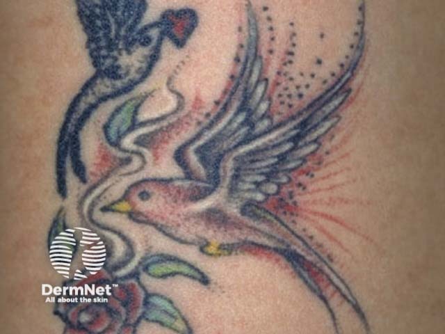
These are usually the result of a delayed hypersensitivity reaction to tattoo pigment and in an attempt to degrade the foreign body material. Again, red pigment is the main cause but it has also been reported with green and blue pigments. Pseudolymphomas caused by tattoo pigment are characteristically plum to red coloured nodules and plaques. They need to be clinically distinguished from cutaneous lymphomas that may be the cause of serious malignant conditions.
The trauma induced by puncturing the skin has been linked to pseudolymphoma which may become generalised over time.
Squamous cell carcinoma, melanoma and leiomyosarcoma have been described in tattoos, particularly in areas of red ink. Keratoacanthoma-like reactions to tattoos have also been described. Although the cause is unknown, it is thought that the ink may include carcinogens. In some cases, the tumours may be due to 'pseudoepitheliomatous hyperplasia', a benign proliferative reaction that mimics skin cancer.
Magnetic resonance imaging (MRI scan) produces a magnetic field that has been reported to induce an electric current within tattoo ink containing iron oxide. Iron oxide is sometimes found in permanent cosmetic ink used to enhance lip lines and eyebrows. A MRI scan results in a minor burn at the tattooed site, ie, painful redness and swelling.
Tattoos are most often treated with Q-switched lasers. In most cases, 5 to 12 treatments are required, at 6 to 8-week intervals. Complete clearance is not always possible.
The laser may be selected according to the colour of the tattoo pigment.
White and yellow pigment appears to be the most difficult to eradicate. Complications may include:
Alternatively, ablative fractional resurfacing can be used.
An AI summary will appear based on your search term using data from all of the topic pages across the entire DermNet site.
Show more