Author: Naomi Ashman, Dermoscopist, Torbay Skin, Auckland, New Zealand; DermNet New Zealand Editor in Chief, Adjunct A/Prof Amanda Oakley, Dermatologist, Hamilton, New Zealand. Copy edited by Gus Mitchell. Created 2019.
Cutaneous squamous cell carcinoma is a common type of keratinocytic or nonmelanoma skin cancer. It is commonly found on sun-exposed areas of skin. It can be invasive and metastasise. It is also known as cutaneoussquamous cell carcinoma, and commonly abbreviated to SCC.
What are the clinical features of squamous cell carcinoma?
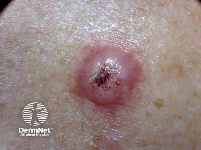
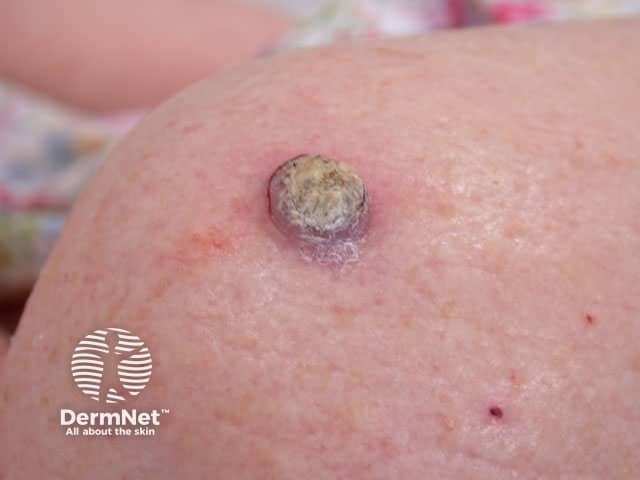
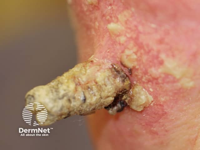
Cutaneous squamous cell carcinoma presents clinically as an enlarging, irregular, keratinous nodule or a firm erythematousplaque that frequently ulcerates.
They usually arise within pre-existing actinic keratosis or intraepidermal carcinoma.
They grow over weeks to months
They may ulcerate
They are often tender or painful
Located on sun-exposed sites, particularly the face, lips, ears, hands, forearms, and lower legs
Size varies from a few millimetres to several centimetres in diameter
They are rarely pigmented.
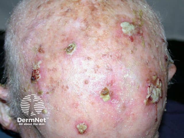
Multiple squamous cell carcinomas on the scalp
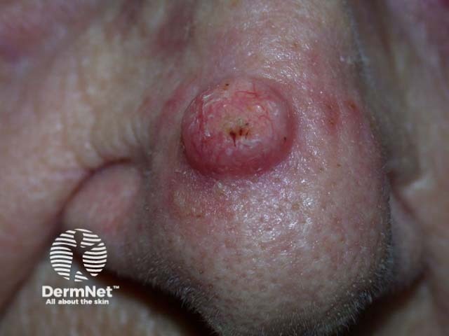
Squamous cell carcinoma of the nose
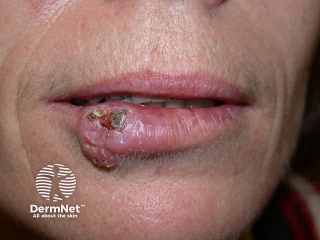
Squamous cell carcinoma of the lip
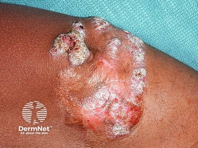
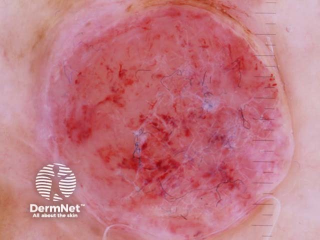
High-risk cutaneous squamous cell carcinoma
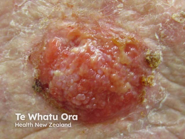
Squamous cell carcinoma
Squamous cell carcinoma
What are the dermoscopic features of squamous cell carcinoma?
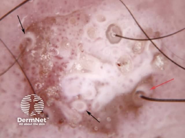
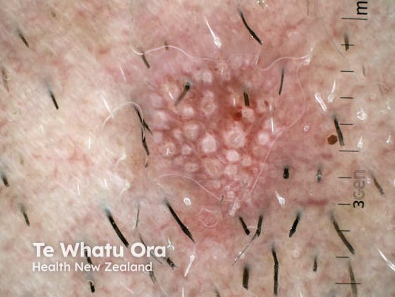
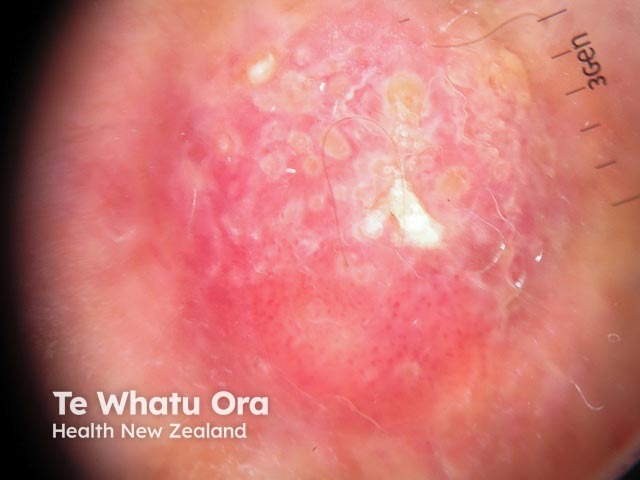
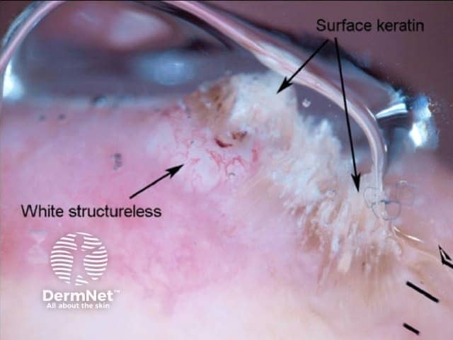
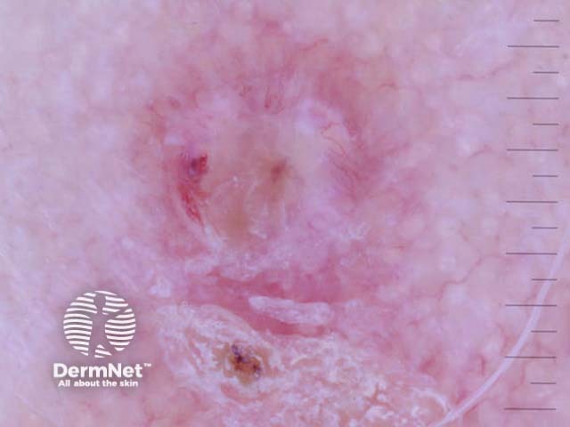
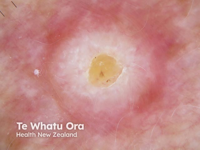
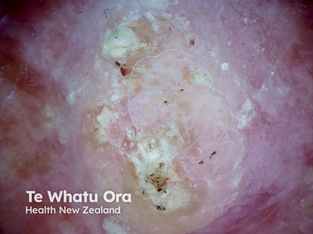
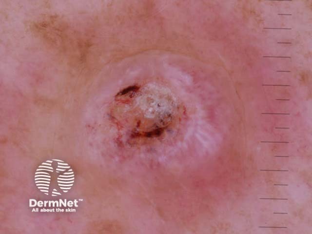
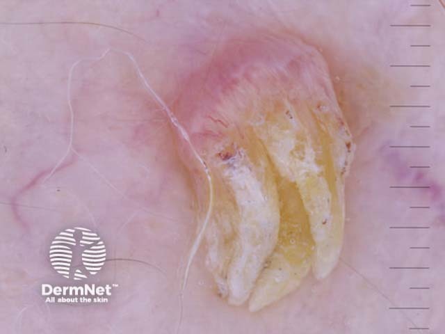
The common dermoscopic features of cutaneous squamous cell carcinoma are:
White circles
White structureless areas
Looped vessels
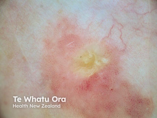
Central keratin
Pink or red background in poorly differentiated or rapidly growing tumours.
White structureless areas and white circles in squamous cell carcinoma dermoscopy
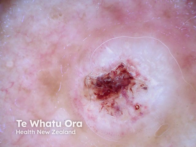
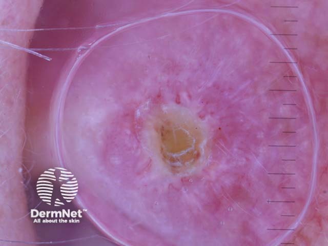
White circles in squamous cell carcinoma dermoscopy
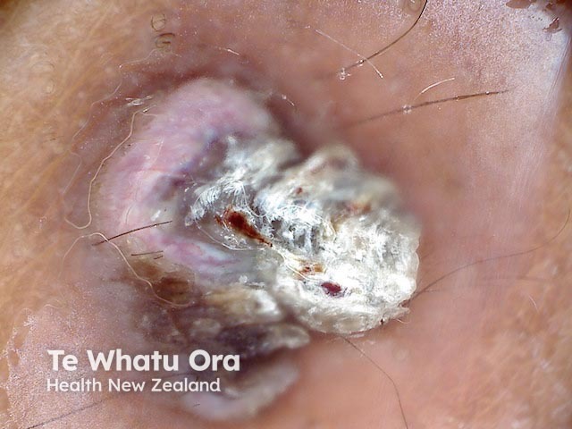
White circles and surface keratin seen in squamous cell carcinoma dermoscopy
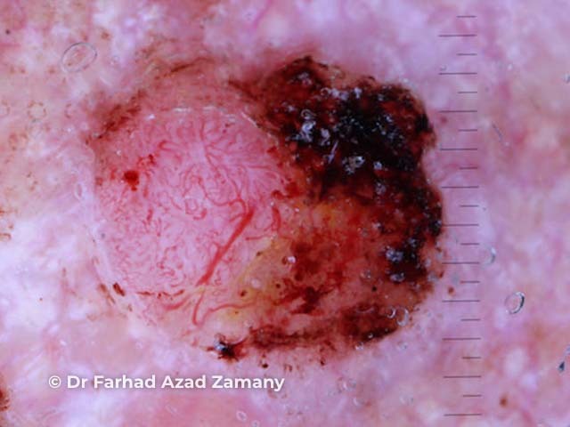
White structures and surface keratin in squamous cell carcinoma dermoscopy
Loop vessels seen in squamous cell carcinoma dermoscopy
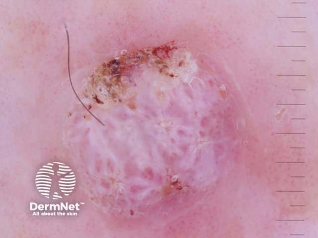
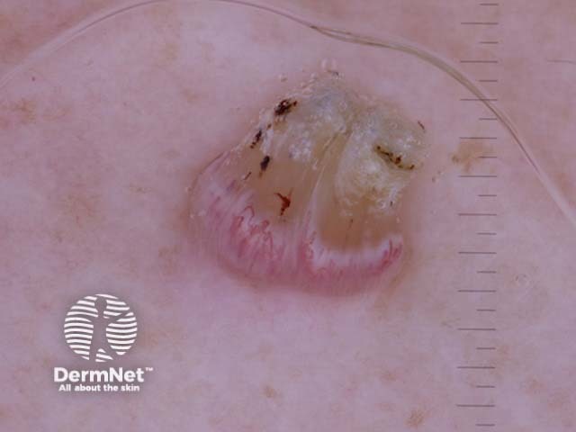
White structureless areas and central keratin in squamous cell carcinoma dermoscopy
White structureless areas and surface keratin in squamous cell carcinoma dermoscopy
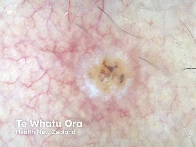
White structureless areas, central keratin and loop vessels in squamous cell carcinoma dermoscopy
Amelanoticmelanoma, Breslow 3.2mm, CL IV dermoscopy
Amelanotic melanoma dermoscopy
What is the histological explanation for squamous cell carcinoma?
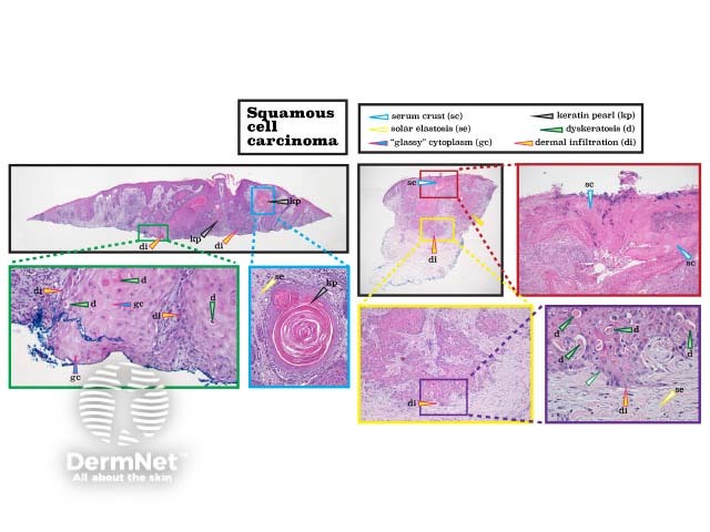
Histologically, there is a proliferation of atypicalkeratinocytes within the dermis.
Histopathology of squamous cell carcinoma
References
Motley R, Kersey P, Lawrence C; British Association of Dermatologists; British Association of Plastic Surgeons; Royal College of Radiologists, Faculty of Clinical Oncology. Multiprofessional guidelines for the management of the patient with primary cutaneous squamous cell carcinoma. Br J Dermatol. 2002;146(1):18–25. doi:10.1046/j.0007-0963.2001.04615.x. PubMed
Parikh SA, Patel VA, Ratner D. Advances in the management of cutaneous squamous cell carcinoma. F1000Prime Reports. 2014;6:70. DOI: 10.12703/P6-70. PubMed Central
Guidelines of care for the management of cutaneous squamous cell carcinoma. Alam, MuradKim, John YS, et al. J Am Acad Derm. DOI: doi.org/10.1016/j.jaad.2017.10.007. Journal