Within the group of cutaneousB celllymphomas, further categorisation includes primary cutaneous diffuse large B cell lymphoma, leg type; primary cutaneous diffuse large B cell lymphoma, other; plasmablastic lymphoma and T cell-rich large B cell lymphoma. The primary cutaneous B cell lymphoma leg type indicates a poor prognostic group typically seen on the lower legs of the elderly and is illustrated in the figures below.
Histology of primary cutaneous diffuse large B cell lymphoma
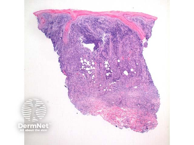
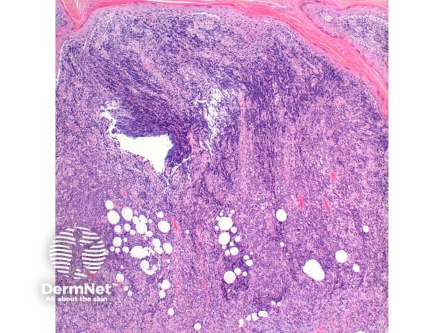
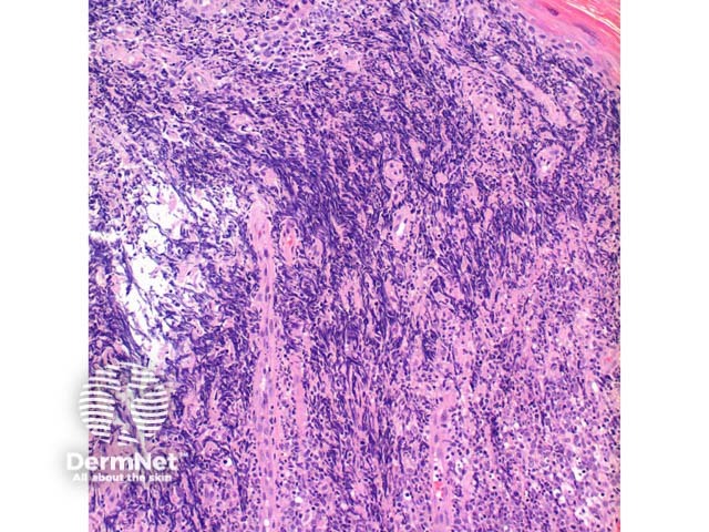
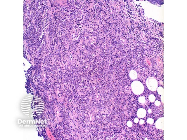
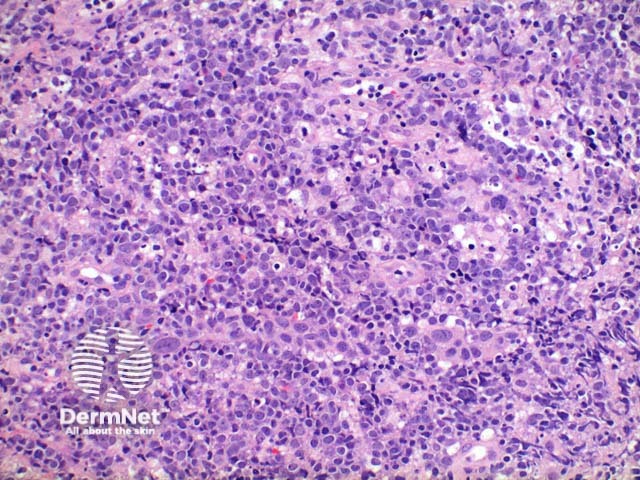
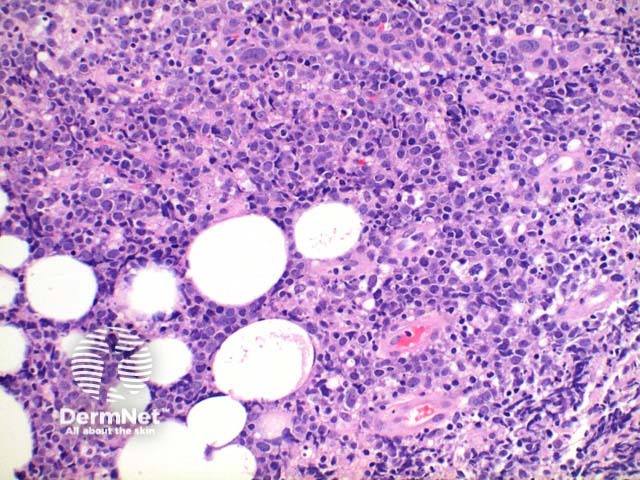
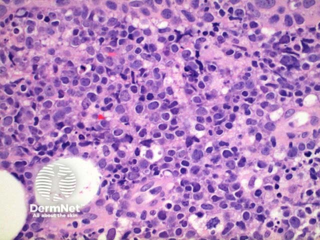
Scanning power view of the histology of primary cutaneous diffuse large B cell lymphoma is of a dense superficial and deep cellularinfiltrate (figure 1). Higher power identifies a dense atypicallymphocyticproliferation, which frequently extends into the subcutaneous tissue (figures 2, 3, 4, 5). Areas of crush artefact are common, and while not specific may be a clue to a malignantlymphoid population (figures 2, 3). The lymphocytes are large centroblast-like and immunoblast-like cells with non-cleaved nuclei and prominent nucleoli (figure 6, 7).
Figure 1
Figure 2
Figure 3
Figure 4
Figure 5
Figure 6
Figure 7
Special stains in primary cutaneous diffuse large B cell lymphoma
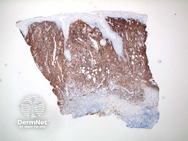
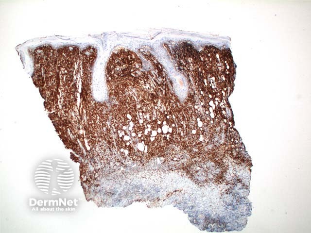
Immunostaining reveals a CD20 positive B (figure 8) cell population with diffuse BCL2 (figure 9) and MUM1 staining. BCL6 expression is variable while CD10 is negative.
Figure 8
Figure 9
Differential diagnosis of primary cutaneous diffuse large B cell lymphoma
Primary cutaneous diffuse large B cell lymphoma, other, refers to those cases where the clinical features, histology and immunohistochemistry do not fit with primary cutaneous diffuse large B cell lymphoma, leg type, while also not consistent with a diagnosis of primary cutaneous follicle centre lymphoma.
References
Kempf W, Sander CA. Classification of cutaneous lymphomas – an update. Histopathology 2010;56:57–70. PubMed
Skin Pathology (2nd edition, 2002). Weedon D
Pathology of the Skin (3rd edition, 2005). McKee PH, J. Calonje JE, Granter SR