Plasma cells are white blood cells produced in the bone marrow and are found in the blood, the skin, and throughout the body. Their function is to make immunoglobulins (antibodies) to fight disease. They are an important part of the immune system.
What is a plasmacytoma?
A plasmacytoma is a tumour made up of abnormal plasma cells. It usually grows within the bone. When it grows in a site other than bone, it is called an extramedullary plasmacytoma.
There can be a single tumour (solitary plasmacytoma) or many tumours (multiple myeloma)
Who gets plasmacytoma?
Plasmacytoma is rare. It affects middle aged and elderly people and is very rare under the age of 30 years. Cutaneous plasmacytoma is very rare.
What is the cause of plasmacytoma?
It is not known what causes plasmacytoma. Radiation, industrial solvents and airborne toxins have been identified as possible risk factors.
How does plasmacytoma present?
Solitary plasmacytoma of bone
Plasmactyoma may cause:
Pain
Pathological fracture
Spinal cord compression
Extramedullary plasmacytoma
Extramedullary plasmacytoma can occur at any site, but 80–90% of extramedullary plasmacytomas are in the head and neck area, particularly within the upper airways and oral cavity. Symptoms may include:
Breathlessness (dyspnoea), coughing up blood (haemoptysis)
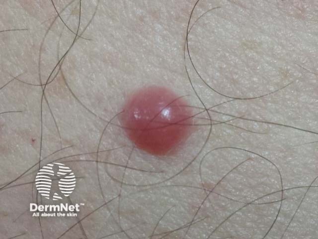
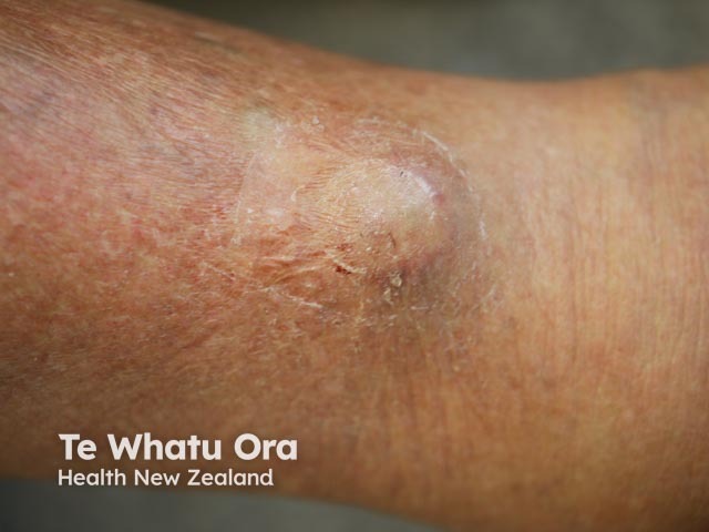
Cutaneous/skin involvement is very rare, accounting for 2–4% of all extramedullary plasmacytomas. They typically present as red nodules or dome shaped plaques, and may ulcerate.
Cutaneous plasmacytoma
How is plasmacytoma diagnosed?
Plasmacytoma is diagnosed by a tissue biopsy or bone marrow biopsy. This shows invasion of the bone or tissue by monoclonal (identical) plasma cells (see cutaneous plasmacytoma pathology). To make a diagnosis of solitary plasmacytoma (of bone or extramedullary site), other plasma cell tumours or multiple myeloma must be excluded.
What is the treatment for plasmacytoma?
Solitary plasmacytoma is treated by radiotherapy or surgery.
What is the prognosis for plasmacytoma?
The prognosis for plasmacytoma depends on whether the lesions are solitary or a sign of multiple myeloma.
Multiple myeloma may later develop in patients with solitary bone plasmacytoma (65–84% at 10 years) or extramedullary sites (11–30% at 10 years).
Cutaneous involvement in patients with known multiple myeloma usually indicates a poor prognosis regardless of treatment.
Multiple myeloma
Normally, plasma cells account for <5% of bone marrow. Plasma cell cancers occur when there is uncontrolled proliferation of these cells. There are two forms:
Multiple myeloma (>10% of bone marrow)
Plasma cell leukaemia (>20% of nucleated peripheral blood cells)
Plasma cell leukaemia is rare and aggressive. It can be primary (the first sign of disease) or secondary to advanced multiple myeloma.
Approximately 10–15% of patients presenting with a solitary extramedullary plasmacytoma and 50–60% of patients presenting with solitary plasmacytoma of the bone will ultimately develop multiple myeloma.
Extramedullary plasmacytomas are seen in around 7% of patients who have multiple myeloma at the time of diagnosis, and a further 6% of patients will go on to develop extramedullary plasmacytomas after being diagnosed with multiple myeloma.
Multiple myeloma presents with the following signs and symptoms.
Bone pain or fractures
Anaemia: weakness, tiredness, breathlessness
Low white cells (leukopenia) may lead to recurrentinfections, eg pneumonia
Thickened blood: confusion, dizziness, stroke
Hypercalcaemia: thirst, dehydration, weakness, bowel and kidney problems
Neurological symptoms: back pain, numbness or weakness
The following tests are used to detect and monitor multiple myeloma.
Full blood count to assess anaemia, low white blood cells and low platelets
Lactic dehydrogenase (LDH), calcium, phosphorus , erythrocyte sedimentation rate (ESR) and C-reactive protein (CRP)—elevated in disseminatedcancer
Creatinine and urea to assess kidney function
Beta-2 microglobulin, which is expressed on the surface of myeloma cells
Serumproteinelectrophoresis with immunofixation, to detect and classify abnormal/monoclonal protein in serum
Serum immunoglobulins, elevated in myeloma
Urinary protein electrophoresis with immunofixation (24-hour urine sample), to detect and classify monoclonal kappa or lambda light chains in urine
Skeletal bone survey to detect bone thinning (osteoporosis, osteopaenia) or holes (lytic lesions) caused by myeloma. MRI, CT and PET scans may also be used in some cases
Treatment
If multiple myeloma is present, it may be treated by:
Treatments may be used individually or in combination, depending on the case details and risk profile.
Prognosis
Predicting prognosis in multiple myeloma is not straightforward, as some patients remain asymptomatic for a number of years while for others the disease is rapidly progressive.
Staging (tumour burden)
Patient characteristics
Disease characteristics
Availability and response to therapy
These estimates of prognosis may be adjusted depending on availability of and response to treatment, patient characteristics, and tumour genetics. Some tumour mutations are associated with more aggressive forms of disease which may shorten survival time.
Patients are categorised as high, medium or standard risk using cytogenetic testing, which is done on the tumour cells to identify geneticabnormalities which may contribute to a more aggressive disease pathway.
The standard and medium risk groups have an estimated median survival of 8–10 years. Patients in whom certain genetic abnormalities are detected are likely to have a shorter survival time, as are patients in whom disease is advanced.
Catarina Araújo, Herlander Marques, José Carlos Fernandes, António Pardal and Celeste Brito. Cutaneous Plasmacytomas Secondary to Nonsecretory Multiple Myeloma. Journal of Dermatology and Clinical Research. SciMed Centrail, Published 6/06/2014
Dimopoulos MA, Moulopoulos LA, Maniatis A, Alexanian R. Solitary plasmacytoma of bone and asymptomatic multiple myeloma. Blood. 2000 Sep 15;96(6):2037–44. Review. PubMed PMID: 10979944.
Dimopoulos MA, Kiamouris C, Moulopoulos LA. Solitary plasmacytoma of bone and extramedullary plasmacytoma. Hematol Oncol Clin North Am. 1999 Dec;13(6):1249–57. Review. PubMed PMID: 10626148.
Muscardin LM, Pulsoni A, Cerroni L. Primary cutaneous plasmacytoma: report of a case with review of the literature. J Am Acad Dermatol. 2000 Nov;43(5 Pt 2):962–5. Review. PubMed PMID: 11044834.
Erdogan BA, Sekercan O, Dursun N, Tatar G, Bora F. Extramedullary plasmacytoma of maxillary sinus. J Craniofac Surg. 2013 Jan;24(1):e85–7. doi: 10.1097/SCS.0b013e31827103e9. PubMed PMID: 23348350.
Requena L. [Specific cutaneous involvement in patients with multiple myeloma. A clinicopathological, immunohistochemical and cytogenetic study of 40 cases]. Actas Dermosifiliogr. 2005 Sep;96(7):424–40. Review. Spanish. PubMed PMID: 16476270.
Yu SC, Chen SU, Lu W, Liu TY, Lin CW. Expression of CD19 and lack of miR-223 distinguish extramedullary plasmacytoma from multiple myeloma. Histopathology. 2011 May;58(6):896–905. doi: 10.1111/j.1365-2559.2011.03793.x. Epub 2011 Mar 14. PubMed PMID: 21401705.
Hu K, Yahalom J. Radiotherapy in the management of plasma cell tumors. Oncology (Williston Park). 2000 Jan;14(1):101–8, 111; discussion 111-2, 115. Review. PubMed PMID: 10680152.
Requena L, Kutzner H, Palmedo G, Calonje E, Requena C, Pérez G, Pastor MA, Sangueza OP. Cutaneous involvement in multiple myeloma: a clinicopathologic, immunohistochemical, and cytogenetic study of 8 cases. Arch Dermatol. 2003 Apr;139(4):475-86. Review. PubMed PMID: 12707095.
Russell SJ, Rajkumar SV. Multiple myeloma and the road to personalised medicine. Lancet Oncol. 2011 Jul;12(7):617–9. doi: 10.1016/S1470-2045(11)70143-7. Epub 2011 Jun 12. PubMed PMID: 21664869.
Multiple Myeloma – The American Cancer Society, accessed 11/09/14
Bladé J, Fernández de Larrea C, Rosiñol L, Cibeira MT, Jiménez R, Powles R. Soft-tissue plasmacytomas in multiple myeloma: incidence, mechanisms of extramedullary spread, and treatment approach. J Clin Oncol. 2011 Oct 1;29(28):3805–12. doi: 10.1200/JCO.2011.34.9290. Epub 2011 Sep 6. Review. PubMed PMID: 21900099.
Fernández de Larrea C, Kyle RA, Durie BG, et al. Plasma cell leukemia: consensus statement on diagnostic requirements, response criteria and treatment recommendations by the International Myeloma Working Group. Leukemia. 2013 Apr;27(4):780–91. doi: 10.1038/leu.2012.336. Epub 2012 Nov 21. Review. PubMed PMID: 23288300; PubMed Central PMCID: PMC4112539.
Varettoni M, Corso A, Pica G, Mangiacavalli S, Pascutto C, Lazzarino M. Incidence, presenting features and outcome of extramedullary disease in multiple myeloma: a longitudinal study on 1003 consecutive patients. Ann Oncol. 2010; 21: 325–330. PubMed