Pityriasis lichenoides et varioliformis acuta pathology
Authors: Dr Achala Liyanage, Dermatology Fellow, Waikato Hospital, Hamilton, New Zealand; Assoc Prof Patrick Emanuel, Dermatopathologist, Auckland, New Zealand. January 2015.
Pityriasis lichenoides et varioliformis acuta (PLEVA) presents with haemorrhagicpapules that resolve to leave varioliform scars. It is usually a self-limitingacutedermatosis. It is also known as Mucha Habermann disease.
Histology of PLEVA
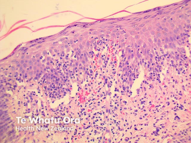
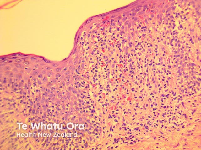
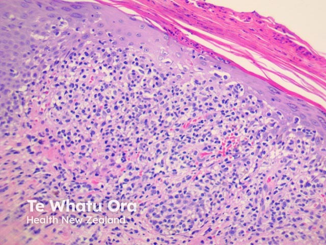
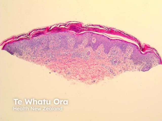
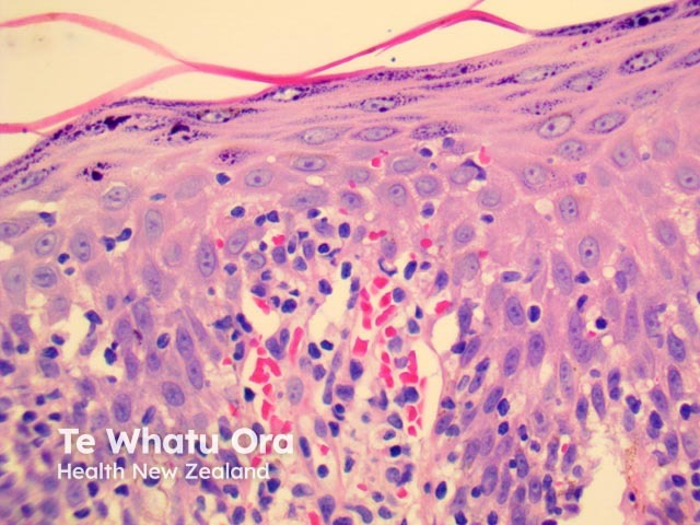
PLEVA has sharply delimited, moderately dense, lymphocyticinfiltrate involving the superficial vascularplexus, which extends in a wedge-shaped pattern to involve the lower dermis (figure 1). The superficial dermis shows a dense lichenoid infiltrate and impressive exocytosis of lymphocytes into the epidermis. The overlying stratum corneum shows parakeratosis which may be confluent and contain collections of neutrophils (figure 2). The epidermis shows pronounced hydropic change and foci of keratinocytenecrosis. Scattered extravasatederythrocytes are seen (figure 3).
Figure 1
Figure 2
Figure 3
Figure 4
Figure 5
Images provided by Dr Duncan Lamont, Waikato Hospital
Special studies in PLEVA
None are generally needed. Immunoperoxidase studies have shown the lymphocytic infiltrate consists of CD8/cytotoxicT cells.
Differential diagnosis of PLEVA
CutaneousT-celllymphoma
Lymphocytic vasculitis
References
Weedon’s Skin Pathology (Third edition, 2010). David Weedon